Jaundice
Historical document, translated for reference. It reflects medical knowledge of the 1920s–30s and is not medical advice.
Summary
This 1930s article from the Great Medical Encyclopedia explores the history, etiology, and pathogenesis of jaundice (icterus), redefining it from an independent disease into a syndrome characterized by hyperbilirubinemia. It discusses mechanical, parenchymal, and hemolytic forms of jaundice, detailing their clinical manifestations, pigment metabolism, and tissue involvement.
Encyclopedia article (1928–1936)
Jaundice, icterus (from Greek icteros, the name of a small yellow bird by looking at which, according to the Romans, one could be cured of jaundice). Another name, now abandoned, is morbus regius (royal disease). Etiology and pathogenesis. Ancient thought under the general name of jaundice united many diseases in which a yellow coloration of the skin and visible mucous membranes was observed in patients. Like "dropsy," "fever," and "dyspnea," since the time of Hippocrates and Galen and right up to the beginning of the 19th century, jaundice was considered an independent disease. Humoral pathology distinguished only two forms of jaundice: benign and malignant fatal—"icterus gravis." However, with the accumulation of new clinical, and especially anatomical and physiological facts, the idea of jaundice as a single and independent disease had to be abandoned, and the clinic has long since looked upon jaundice as a symptom of many and very diverse pathological conditions. From the modern point of view, jaundice is a symptom manifested by a yellow coloration of the skin and visible mucous membranes by the bile pigment bilirubin as a result of its increased content in the blood plasma. At first glance, it may seem that the modern clinic has narrowed the concept of jaundice and reduced it to only one phenomenon, namely, an increased bilirubin content in the blood, or hyperbilirubinemia. True, more precise methods have shown that normal blood serum also contains a certain, quite definite amount of bilirubin and that the varying degree of jaundice depends on the degree of accumulation of this pigment in the blood. Thus, proceeding from this sign, one can speak 1) of latent and initial forms of jaundice still without coloration of the skin and mucous membranes, 2) of jaundice in which the skin is colored, but bilirubin does not penetrate into the urine and only urobilin is contained in it, and finally 3) of expressed jaundice, when hyperbilirubinemia is so significant that the visible yellow coloration of the integuments is also expressed more sharply and bilirubin is easily determined in the urine. From this it is seen that at the heart of jaundice lies only an intensification of the physiological process, i.e., the content of bilirubin in the blood. Thus, at present all types of jaundice are united by one sign, namely, hyperbilirubinemia. However, along with this, the same more refined methodology made it possible to establish a whole series of new facts concerning pigment metabolism. The relationship between blood hemoglobin, bilirubin, and urine urobilin and fecal urobilinogen has been established. If the question of the site of formation of bilirubin and urobilin cannot be considered completely resolved, one cannot fail to recognize the paramount role of the hepatic cell in the excretion of these pigments. At the same time, the possibility of the appearance of hyperbilirubinemia with jaundice without the direct participation of the liver with enhanced erythrocyte destruction (hemolytic jaundice) has been proven. Furthermore, certain forms of jaundice reveal definite, regular relations between the content of bilirubin and urine urobilin and fecal urobilinogen (in mechanical jaundice). In retention jaundice, as has been clarified at present, there is a retention in the blood, besides pigments, of other constituents of bile: bile acids and cholesterol, while in other cases a dissociated retention of bile elements is possible, i.e., bile pigments or bile acids can be retained in the organism in an isolated manner (dissociated jaundice of French authors). If to all that has been said one adds that often in combination with the icteric skin coloration a whole series of other clinical symptoms closely related to jaundice are observed, then at present the concept of jaundice can again be expanded to a syndrome, a symptom complex, or even to a very complex special pathological process. Modern clinics speak not of jaundice, but of jaundices, and each case requires a careful individual clinical analysis. In some cases, jaundice is a concomitant symptom, and its pathogenesis appears relatively simple and elucidated, e.g., obstructive, or mechanical, or retention jaundice upon blockage of the common bile duct by a stone or upon its compression by a cancer developing in the head of the pancreas. In other cases, the mechanism of jaundice is more complex, with the lesion of the hepatic cell itself playing the main role (hepatic, parenchymal jaundice). Finally, the third form of jaundice—hemolytic (acholuric, dynamic)—occurs without the direct participation of the liver as a result of the enhanced destruction of erythrocytes. At present, all three forms of jaundice are described in the corresponding diseases (jaundice in gallstone disease, in liver diseases, in various infections, in poisoning, etc.). However, even now such relatively insufficiently studied disease forms are observed in which this symptom occupies a prominent position in the entire clinical picture and is its main clinical characteristic. Such jaundices, despite the fact that their etiology is sometimes known (infection, toxin) and their pathological anatomy is clarified (hepatitis, angiocholitis), are described under the name of "jaundices" as special independent symptom complexes. The boundaries of these forms are extremely wide—from simple, or in previous terminology, catarrhal jaundice to icterus gravis, or to acute yellow atrophy of the liver. In the pathogenesis of these jaundices, the lesion of the hepatic cell itself stands in first place. The latter participates to a greater or lesser extent in all jaundices; according to one theory, it can either lose the ability to produce bile elements (acholia) or produce them in abundant quantity (pleiochromia); according to another theory, it either retains urobilin or bilirubin formed outside the liver (in the reticuloendothelial system) or excretes them in an increased quantity. The yellow coloration of the skin and mucous membranes is a consequence of the pathological increase of bilirubin in the blood. Depending on the intensity of hyperbilirubinemia, the brightness of the skin coloration also varies—from a light lemon-yellow shade to orange-yellow and green or olive-yellow color, which served as the occasion for the name melas icterus (black jaundice). The distribution of the pigment occurs unevenly. The mucous membranes are colored first of all (conjunctiva, lower surface of the tongue, palate), then skin folds on the face, the skin around the nose and mouth, palms, and soles. Later, the entire skin is colored. In mild cases, the matter is limited to the coloration of the conjunctivae; then they speak of subicterus. The coloration of the skin is proportional to the degree of hyperbilirubinemia and the severity of jaundice. Hyperbilirubinemia is the only constant sign of jaundice. All other symptoms, e.g., the retention in the blood of bile acids, etc., are not always observed; they vary depending on the clinical form of jaundice. Hyperbilirubinemia serves as the unifying moment for all types of jaundice. The retention of bilirubin in the organism besides the special coloration of the integuments does not cause any toxic disorders (see also Bile - bile pigments). Clinical study of hemolytic jaundices, in which the increased bilirubin content in the blood persists for months without the parallel retention of other elements of bile, makes it possible to consider the toxicity of bilirubin minimal. Bilirubin stains not only the skin, but also almost all tissues of the organism. Serous membranes are impregnated with pigment first of all, then connective fibrous tissue, later epithelial cells, further bone tissue; cartilages are not stained at all. Normal secretions of the organism—sweat, saliva, tears, milk—contain little bilirubin even in intense jaundice. But in the presence of an inflammatory process in any of the secreting organs, the secretion produced by the organ contains a significant amount of pigment. In the same way, bilirubin is also contained in pathological accumulations (ascites, exudate, etc.). The pigment does not penetrate into the cerebrospinal fluid due to the impermeability of the meninges to coloring substances. If the liver is healthy and the bile passages are patent, bilirubin accumulating in the blood in excess is removed through the liver into the intestine; upon closure of this pathway, bilirubin is excreted through the kidneys with the urine. Since jaundice most often deals with a lesion of the liver or its pathways, urine usually changes its color fairly soon and acquires a characteristic dark coloration. True, bilirubin appears in the urine not immediately, but only when it reaches a sufficient concentration in the blood serum and is determined by the direct Giemans van den Berg reaction; with weakly expressed hyperbilirubinemia determined by the indirect reaction, another pigment—urobilin—is determined in the urine. From here comes the previous name of weakly expressed jaundice—"urobilinic." Urobilin is contained in the urine not only in hemolytic jaundice, but also at the beginning of the development of other jaundices, while bilirubinemia is weak and does not reach that degree of concentration at which bilirubin also appears in the urine. The impregnation of the skin with pigment requires a certain time. In experimental ligation of the bile duct, bilirubinemia increases after 24 hours, while the skin is colored after a few days. Clinical observations show that upon blockage of the common bile duct, jaundice develops 5-8 days later. In certain poisonings, e.g., with toluylenediamine, jaundice is outlined already after a day, although it reaches a maximum on the third day. The pathogenesis of jaundice is complex.
Jaundice accompanies a wide variety of diseases (diseases of the liver and gall bladder, infectious and intoxication diseases, anemias, diseases of the spleen, etc.), and sometimes also physiological conditions (icterus menstrualis, gravidarum, neonatorum). Nevertheless, all types of jaundice can currently be reduced to three main pathogenetic forms: 1) mechanical, or retention jaundice, 2) parenchymal, or hepatic jaundice, and 3) hemolytic, or dynamic jaundice. I. Mechanical jaundice (see separate table, Fig. 1) has been studied the most. Formerly, the majority of cases of hepatic jaundice (e.g., icterus simplex) were also referred to this group; now, only those diseases in which there are anatomically demonstrable Figure 1. Obstructive jaundice (cancer of the pancreas) are classified as mechanical jaundice.
\
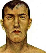
Figure 2. Simple, 'catarrhal' jaundice. Figure 3. Hemolytic jaundice. To the article Jaundice. B. L1. E. *"1ST ,' 1C н * * ТИR0«' obstacles to the passage of bile through the bile ducts. In the presence of a mechanical obstacle, above it forms, due to increased pressure within the ducts with continued bile secretion, dilation of the bile ducts, which can reach very significant sizes. Secretion of bile continues, although its amount decreases and at times there is impoverishment of it in pigment. According to the research of Eppinger, stretching of the bile capillaries can end in their rupture, as a result of which communication is established between them and the lymph spaces and blood vessels. The bile that has flowed into these spaces freely enters the general bloodstream. In mechanical J. not only bilirubin but also other ingredients of bile-bile acids, cholesterol-are absorbed into the blood. As a result, in addition to hyperbilirubinemia, hypercholesterolemia develops and according to the terminology of French authors-"cholemia" (accumulation in the blood of bile acids). The combination of the listed symptoms creates the picture of cholemia. The main role in the development of the picture of cholemia belongs to the bile acids, the accumulation of which causes a series of toxic vascular-nerve phenomena. In addition to the symptoms caused by the retention of bile pigments and acids in the body, mechanical J. is characterized by phenomena caused by the absence of bile in the intestine (see below). In almost all cases of mechanical J. the liver increases in volume, acquires a dark green color and becomes firm in consistency. The surface of the liver on cross-section is dotted with dark spots ranging in size from a pinhead to a lentil; in the center of the spots there are depressions, less intensely colored in yellow than the peripheral parts. These spots are the result of the so-called bile apoplexies due to the retention of bile and increased pressure within the bile ducts. The ducts themselves are dilated and in severe cases are either ulcerated or thickened and filled with colorless mucus with an admixture of a small amount of bile. Liver cells, especially those located around the central vein, are imbued with bile pigment, often depositing in the form of clumps and lumps. This pigment also often stains the nucleus of the cells. If the obstruction of the bile ducts continues for a long time and is complicated by infection, then mechanical jaundice ends either with diffuse intraparenchymal sclerosis with atrophy of many liver lobules and thickening of the interstitial tissue (secondary cirrhosis of the liver) or it is complicated by purulent angiocholitis with small abscesses around the bile ducts. In addition to the liver, the tissue of the kidneys is also impregnated with the coloring substance of bile. From the outside the kidneys appear almost olive-green in color, and on the surface of the cross-section-yellow-red; in the epithelium and in the lumens of the urinary tubules numerous pigment clumps are noticed. A long-lasting mechanical J. gives a picture of a degenerative process in the hepatic epithelium, and along with secondary cirrhosis, nephrosis-nephritis can develop. The pancreas undergoes special changes in cancer of its head, compressing the bile duct and causing obstructive jaundice, but also in mechanical J. from other causes in the pancreas there is also an imbibition of its cells with bile pigment and the development of sclerosis with proliferation of interstitial tissue (chronic pancreatitis). P. The pathogenesis of parenchymatous jaundice is much more complicated, an example of which can be icterus simplex (see separate table, fig. 2). Previously, to explain this disease, mechanical causes in the form of swelling of the mucous membrane at the point of entry of the ductus choledochi into the duodenum or in the form of a mucous plug blocking the duct (Virchow) were sought. However, the inconsistency of such interpretations with path.-anat. data forced clinicians to seek other explanations. One of the first theories was the theory of the so-called 'Suppressionicterus'; in general outline it was formulated as early as Morgagni. According to it, the diseased liver ceases to secrete from the blood already preformed, bile constituent elements found in it. This theory has not lost its significance to this day, especially in connection with the fact that for certain forms of J. it can be considered proven that bilirubin can be formed from blood pigment outside the liver in the cells of the ret.-end. system. This extrahepatic formation of bilirubin is acceptable for explaining hemolytic J., and possibly also for some hepatic J.; in these cases the diseased liver cell loses the ability to excrete this pigment. In 1881 Stadelmann, working with icterogenic poisons (toluenediamine, phosphorus), proved that under their influence a larger amount of bile richer in pigment is secreted. He raised the question of polycholia and polychromia, or pleiochromia as a pathogenetic moment of J.; according to him, concentrated bile itself is a mechanical obstacle to its passage. Subsequently Eppinger confirmed the significance of pleiochromia by finding in it in the liver bile thrombi blocking the bile capillaries. One cannot deny the existence of pleiochromia. However, most likely it does not play the role of a primary pathogenetic moment, but is one of the manifestations of increased hemolysis in general and hemolytic J. in particular. Minkowski, not being satisfied with the theory of pleiochromia, proposed the hypothesis of 'parapedesis of bile'. In the normal liver bile is secreted and excreted by liver cells only in one definite direction-to the bile ducts, while sugar and urea enter only the vascular bed. In path. cases the bile produced by liver cells begins to enter by a second path. Similarly, the view of Pick on 'paracholia'. Libermeyer introduced the term 'acatastatic J.'; it arises in his opinion due to the loss by the liver cell of the ability to prevent the diffusion of bile into the lymphatic pathways. All these hypotheses have only historical interest. True, even now the exact mechanism of development of hepatic J. is unknown, but it has been established that the function of the liver cell as a whole suffers and that in many cases the functional disorders correspond to the picture of anat. destruction. Observations on infectious J. showed that the mildest cases are accompanied by significant disturbances of the glycogenic function of the liver, nitrogenous, water and salt metabolism. Brulé and Lemiere described 'dissociated J.', in which in the body either bile pigments or bile acids are isolatedly retained. They drew a parallel with kidney lesions and dissociated retention of NaCl and N and saw in dissociated J. a new confirmation of the correctness of the theory of parenchymatous jaundice. Fiessinger and Lyon-Coen found dissociated J. in phosphorus poisoning, Lemière and Brulé- under the action of hepatotoxic sera. Some German authors, in particular Umber, refute the teaching on dissociated J. The main objection is the indication of the inaccuracy of the method for determining bile acids by Heycraft, which was used by Lemière and Brulé. J., observed during many infectious diseases, often depends on damage to liver cells by microbes or, rather, their toxins, which, being excreted from the body by this path, must pass through the liver cell to enter the bile ducts. Such a transient intercurrent J. French authors compare with albuminurias complicating acute infections. Eppinger repeatedly performed histological examination of the liver in icterus simplex, finding b. or m. widespread necroses of the parenchyma. Due to the close connection of liver cells with capillaries, the latter are involved in the degenerative process and are destroyed; as a result, communication is formed between the bile capillaries and lymph spaces, similar to that which occurs in mechanical J. Between icterus simplex and icterus gravis, characterized by acute yellow atrophy of the liver, there are numerous intermediate forms. Moreover, icterus simplex sometimes passes into acute atrophy of the liver. The latter often begins with the same symptoms that are characteristic of simple J. All these clin. facts prove that at the basis of both diseases lies the same pathological process of varying intensity, i.e., a degenerative parenchymatous process affecting the liver tissue, specifically liver cells. III. Hemolytic J. represents a special pathological form (see Hemolytic jaundice). If the question of the dependence of J. on hemolysis does not present doubts at the present time, then in the pathogenesis of this form of J. there are still many controversial and unclear points. The most important is the question of how the destruction of erythrocytes leads to jaundice. As for the specific question of the transition of Hb into bilirubin, at the present time it is necessary to consider b. or m. probable the extrahepatic formation of this pigment with the participation of cells of the ret.-end. system (Aschoff, Mac Nee, Mann and Magath). It is assumed that in the hemolytic form of J."
(so-called anhepatogenic) liver cell remains unaffected; it simply does not have time to secrete the bilirubin formed in excess, as a result of which hyperbilirubinemia occurs with subsequent jaundice (see separate table, fig. 3). A significant difference and feature of most cases of hemolytic J. is the delay of only bile pigments without simultaneous delay of other bile components: bile acids and cholesterol. Unlike hepatic and mechanical J., in this form toxic phenomena (bradycardia, skin itching, etc.) are only rarely observed. However, besides these clearly defined forms, there are some that do not fit into any of the categories; these are mixed forms. Thus, J. in so-called hypertrophic cirrhosis of the liver has features of both parenchymatous and hemolytic J. Eppinger distinguishes 4 pathogenetic groups of J. and considers it possible to distinguish an even greater number. Thus, the last word on the pathogenesis of J. has not yet been said. - Regarding the etiology of J., one can only speak when describing each clinical form separately. Clinical forms. By mechanical J. is meant all cases in which obstacles, located either inside or outside the bile ducts, hinder the outflow of bile into the duodenum. Internal obstacles include gallstones, ascarids crawling into the bile duct, echinococcal cysts, scars after ulcers of the mucous membrane, and congenital deformities of the bile passages. External obstacles include cancer of the head of the pancreas, cancer of the Vaterian papilla, areas of the porta hepatis, tumors of lymph glands in the hepatic portal areas, duodenal ulcer, perivisceral scar processes in the area adjacent to the common bile duct, aneurysms of the abdominal aorta, hepatic and superior mesenteric arteries, finally tumors of the right kidney, accumulation of fecal masses in the transverse colon, and pregnancy. Depending on whether the common bile duct or one of the hepatic ducts is blocked, complete or incomplete obstruction of the bile passages is observed. With complete obstruction of the common bile duct, there is enormous dilation of the bile passages, including the gallbladder, which takes the form of a stretched bag and can be palpated as an elastic tumor (Courvoisier's symptom, fig. 1). An increase in the gallbladder is absent in cases where it has lost its elasticity due to previous inflammatory processes. The volume of the liver increases. Accumulation of bilirubin in the blood leads to progressive jaundice; the amount of bilirubin in the blood can exceed the norm 20 times. Accumulation of bile acids in the blood causes a number of toxic symptoms. Bradycria appears, the pulse drops to 50-40 beats per minute. Frerichs observed in one case a pulse equal to 21 per minute. With prolonged jaundice, bradycardia gradually decreases and even disappears completely. Blood pressure is lowered. On auscultation of the heart, a systolic murmur is often determined.
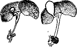
Figure 1. Courvoisier's symptom: on the left, with obstruction of the d. choledochus by a stone, the gallbladder is not enlarged; on the right, with compression of the d. choledochus by a tumor - a large gallbladder.
from the blood side, an increase in the osmotic resistance of erythrocytes is noted. From the nervous system side, general asthenia, headaches, depression, muscle fatigue, sometimes increased neuromuscular excitability, and decreased tendon reflexes are observed. Particularly distressing is the skin itching, which intensifies at night. It occurs, according to Frerichs, in 20% of cases. The itching more often spreads over the entire skin, sometimes localizing only on the palms, soles, and between the fingers of the hands and feet. The itching is explained by irritation of the skin nerve endings by bile acids. There is no direct relationship between the degree of jaundice and itching. Some clinicians point out that in cancer patients, itching is much more pronounced than in patients with gallstones. Sometimes itching precedes the appearance of jaundice. Due to itching, the skin often has scratches that can become infected. In addition, urticaria, herpes circinatus, is sometimes observed. In rare cases, xanthelasma occurs - yellow spots, first localized on the eyelids, and then on other areas of the skin and mucous membranes. Rare cases include xanthopsia, the exact causes of which are still unknown. It is assumed that it depends on toxic changes in the retina; other authors explain xanthopsia by the impregnation of the vitreous body with bile pigments that absorb blue and violet rays. From the intestine side, typical symptoms are observed. With complete obstruction, the stool is acholic, has a clayey gray appearance. The reaction to stercobilin is negative. The stool contains a large amount of fatty acids. Neutral fat is absent in the stool, because it is broken down by pancreatic juice into fatty acids even in the absence of bile. The presence of neutral fat in the stool indicates damage to the pancreas. alimentary lipemia is absent in mechanical jaundice. This forms the basis for the diagnostic test for 'hemokonia,' proposed by Lemierre and Brulé. These authors described in 1910 a simple method that directly allows one to judge the passage of bile acids into the intestine. It is known that the breakdown of fats in the intestine and their absorption from the intestine into the blood occurs under the influence of bile acids, but the determination of alimentary lipemia by chemical means is a difficult and lengthy matter. The test for hemokonia, however, is very simple. If a healthy subject is given a piece of bread with 50 g of oil and after 2 hours a drop of his blood is examined under the ultramicroscope, one can see on a black background plasma formations with shiny grains exhibiting Brownian movement; these are the so-called hemokonia. It has been proven that hemokonia are small particles of fat absorbed in the intestine and entering the general bloodstream. Lemierre and Brulé showed that in patients with complete obstruction of the common bile duct, hemokonia do not appear in the blood; with insufficient bile entering the intestine, hemokonia are found only in small amounts. The duodenal juice in complete mechanical J. is colorless, as it contains no pigments. The Melzer-Lyon test is negative: in response to the introduction of magnesium sulfate into the intestine, bile does not enter the intestine. Valuable diagnostic data is provided by the study of urine. In it, bile pigments (Gmelin's, Sal'kovsky's, Marshal's tests) and bile acids (Gay's, Pettenkofer's reaction, stalagmometry) are found. Urobilin is always absent in complete J. Its appearance indicates restoration, even if partial, of the patency of bile into the intestine (fig. 2). The absence of urobilin in urine with complete obstruction of the common bile duct is explained by F. Müller's theory, according to which
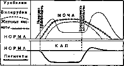
Figure 2. Incomplete retention jaundice.
urobilin is formed in the intestine from bilirubin, is absorbed into the blood by the portal system, and is then again retained by the liver. For urobilinuria to occur, two conditions are necessary: the patency of bile into the intestine and the insufficiency of liver function in terms of its retention of urobilin. With complete obstruction, the first condition is absent, and therefore urobilinuria is also absent (Fig. 3). Mueller's theory fully corresponds to the facts observed clinically. The old theories of urobilin formation in the blood, in the kidneys (Brule and Garban), and the liver theory of Hayem and Tissier have been abandoned. At present, Mueller's theory competes with the hypothesis of Gilbert and Herscher, who believe that urobilin is formed in all cells of the body; according to them, both urobilin and bilirubin have certain thresholds for excretion by the kidneys, with the threshold for urobilin excretion being much lower than that of bilirubin. The same is indicated by experiments of direct introduction of urobilin and bilirubin into the blood (Fromhold, Nersesov). This difference is used to explain the replacement of urobilinuria with bilirubinemia under certain clinical conditions. In recent years, F. Mueller's theory has received full confirmation in the impeccable experimental works of Mac Master and his colleagues. The numerous functions of the liver in mechanical jaundice, as shown by functional tests, remain untouched for a long time; in other words, the liver parenchyma, at least for some time, does not suffer. The course of mechanical jaundice depends on the underlying disease that caused it. Sometimes jaundice ends spontaneously within a few days, sometimes it persists for several months, and sometimes it takes an intermittent course. Parenchymatous jaundice is characteristic of infectious and toxic diseases in which the liver parenchyma is affected. The liver is an excretory organ with antitoxic properties: various infectious agents and toxins are partly detoxified and partly excreted by it. Infectious jaundice can be divided into primary and secondary. By the route of infection, jaundice is divided into ascending infectious jaundice (from the intestine) and descending (from the blood). Secondary jaundices are observed during acute infectious diseases: typhoid fever, paratyphoid fevers, pneumonia, sepsis, etc. They are caused by the agents of the underlying disease that produce infectious hepatitis. Secondary ascending infection from the intestine is of great clinical importance; the main role here belongs to the coli group of bacteria (see Infectious jaundice). The most common form of parenchymatous jaundice, icterus simplex, is also predominantly an infectious disease. The old names—icterus benignus, icterus catarrhalis, which are still sometimes encountered—are deeply incorrect. The first name too often does not correspond to the prognosis, the second does not coincide with the pathogenesis. Mild forms of simple jaundice begin without general symptoms, with the appearance of mild subicterus. Very often jaundice appears after dietary errors, which are followed by diarrhea or constipation. The disease can end in a few days. Much more often the disease begins with general symptoms—chills, a feeling of fatigue and depression, sometimes slight pains in the bones. Appetite is absent, aversion to meat and fats appears, nausea, sometimes pain in the epigastric region and right hypochondrium. The tongue is coated, and from the side of the intestine, diarrhea or, conversely, persistent constipation is observed. Temperature reaches 38-38.5°. On the 3rd-4th day, the urine takes on a dark tint, urobilin is found in it, and later—bile pigments and acids. On the 5th-6th day, yellowish discoloration of the skin appears [see separate table (pp. 15-16), Fig. 2]. From this moment on, jaundice gradually increases. With the appearance of jaundice, the temperature often falls. Bradycardia appears, sometimes—skin itching. The stool becomes discolored. The liver is palpable. Sometimes the spleen enlarges. Functional tests indicate a deep disturbance of liver activity. alimentary glycosuria (ChauHard), levulosuria and galactosuria (Bauer), increase in lipase in the blood (Whippie), changes in nitrogen metabolism (Lasane and M. Weil), increase in blood and enhanced excretion with urine of amino acids (Labbe and Bit, Eppinger), increase in blood of residual nitrogen (Oddo), disturbance in the excretion of glucuronic acid (Gauthier, Clogne and Fissenger), and finally disturbance in water-salt metabolism (Widal and others) have been described. These changes are observed not only in severe but also in mild cases. On the 12th-15th day, jaundice decreases, the urine lightens, the stool becomes colored. Recovery occurs slowly. Even after the disappearance of jaundice, general weakness and weight loss remain, and only after several weeks does restitutio ad integrum occur. In some cases, jaundice drags on for several weeks. In others, it begins to progress rapidly, passes into icterus gravis, and patients die; at the autopsy table, a typical picture of acute yellow atrophy of the liver is found. Finally, in some cases, a tendency to relapses is revealed, which ultimately leads to the formation of persistent hepatitis and even cirrhosis of the liver. The patho-anatomical changes in icterus simplex are little studied, since this form either ends in recovery or passes into acute yellow atrophy of the liver. Eppinger collected a small amount of material, noting the phenomena of degeneration of the liver parenchyma. Another form of parenchymatous jaundice—toxic jaundice—is caused by poisoning with mushrooms, phosphorus, salvarsan, chloroform, picric acid, chlorobenzene, trinitrotoluene and other poisons acting on the liver cell. These jaundices occupy a special chapter in the clinic of poisonings. The reason for this frequent involvement of the liver in suffering is that the liver is the main assimilating and dissimilating gland. Poisons are retained in it, even those that have passed through the portal circulation; here they are transformed, partly destroyed, and partly excreted. Some poisons (arsenic, phosphorus) give a typical picture of liver, parenchymatous jaundice, while others (arsine, acting hemolytically on the blood) give characteristic signs of hemolytic jaundice, and in other poisonings mixed pictures are obtained. The most common is salvarsan jaundice, which arises after prolonged treatment with arsphenamine. Cases of salvarsan jaundice that occurred during amebic dysentery treated with salvarsan have been described; but more often jaundice is observed in the treatment of syphilis. Millian and some other authors considered salvarsan jaundice as a specific luetic. This opinion is incorrect. It is necessary to distinguish between syphilitic jaundice on the basis of specific hepatitis, sometimes observed in the secondary period, from salvarsan jaundice, which arises due to the toxic effect of arsphenamine on the liver cell. Salvarsan jaundice develops without prodromal phenomena; in its course it resembles icterus simplex. In addition to the yellow discoloration of the skin, complete or partial discoloration of the stool, bilirubinuria, and enlargement of the liver are observed. The spleen sometimes enlarges. Liver functions are disturbed and are fully restored only several weeks or even months after the disappearance of jaundice. Jaundice lasts 2-4 weeks, then gradually disappears. Sometimes jaundice takes the character of icterus gravis, and patients die from phenomena of cholemia hemorrhages. Picric jaundice was studied in detail during the last war, when mass poisonings with picric acid for simulation purposes were observed. At first, this form was considered false jaundice, assuming that the discoloration of the skin and mucous membranes is caused not by bilirubin but by picric acid. Later it was proved that the latter is a true liver poison acting on the parenchyma and causing true jaundice. Some authors (Castaigne, Merklen) still believe that the discoloration of the skin after poisoning with picric acid depends mainly on the poison and only to a small extent on bilirubin. However, Brule, Javillier and Baeckeroot showed that after large doses of picric acid, true severe jaundice (icterus gravis) can develop. Picric jaundice is recognized on the basis of finding the specified poison in the blood serum. Jaundice after chloroform anesthesia has practical significance. It usually occurs after prolonged anesthesia. Predisposed are patients with liver diseases (gallstone disease, cholecystitis, hepatitis, etc.). In some patients, the matter is limited to subicterus, in others it reaches icterus gravis with phenomena of cholemia. After ether anesthesia, jaundice is never observed, although cases of increased bilirubin in the blood have been noted. After abuse of alcoholic beverages, alcoholic jaundice often develops (it is more common in alcoholics with chronic hepatitis). Poisoning with mushrooms is sometimes accompanied by jaundice. The nature of jaundice in these cases is still insufficiently studied, but apparently it is a matter of liver parenchyma damage.
Eppinger in one of his cases observed pleochromia, in another - fatty degeneration of the liver, similar to phosphorus poisoning. Phosphorus poisoning simulates the picture of acute yellow atrophy of the liver. In the most severe cases, patients die before the appearance of J. The latter develops on the 4-5th day and proceeds as icterus gravis. Cholemia and bleeding are characteristic.-Besides the listed poisons causing parenchymal J., there is still a whole series of icterogenic substances which, however, cause not parenchymal, but hemolytic J. (arsine). Jaundices with unclear pathogenesis include icterus menstrualis, neonatorum and ex emotione (see below). Menstrual J. was described by Frerichs and Senator (Senator). Apparently it belongs to hemolytic J. and is connected with the formation in the blood of special hemolysins. Similar J. sometimes appear in severe anemias associated with large blood losses (metrorrhagias, hematurias in carcinoma of the bladder-Widal, Joltrain). In the literature there are descriptions of cases of emotional J. that developed after mental shocks. Such J. are explained by spasm of the bile ducts and in particular by spasm of the sphincter described by Oddi and located in the distal end of d. choledochi. The spasm apparently arises due to irritation of the autonomic nervous system (p. splanchnici), which leads (according to experiments by Doyon) to vigorous contraction of the bile ducts. Some clinicians doubt the possibility of the existence of emotive J. and consider the psychological factor a mere coincidence; as an argument against spasm, Chauffard points to the absence of discoloration of the feces in them or to the late disappearance of urobilinogen in the excretions. However, at present it is difficult to deny the influence of the autonomic nervous system on both the lumen of the bile ducts and the liver cell itself.-The prognosis is connected with the cause of J. In mechanical J. it is favorable if the obstacle is removable. In general, the prognosis in J. is fully characterized by the words of Trousseau: "We know very well how J. begins, but we never know how it will end". Prevention and treatment of J. With the progress of knowledge of the pathogenesis of various clinical forms of J., in many cases their etiological therapy can be established. Among mechanical J., some can possibly be prevented by appropriate dietary and general regimen and treatment with mineral waters (gallstone disease), and many of them are radically cured by surgery (removal of stones from d. choledochus) or palliatively (formation of anastomosis with the gallbladder and biliary tract-in carcinoma of the head of pancreas or in other cases by drainage of the biliary ducts). In hemolytic J., prevention of infections (syphilis, malaria, etc.) and intoxications (professional poisons) is important in terms of prevention; in the treatment of hemolytic J., removal of the spleen (splenectomy) often brings benefit. In liver J., the cause can often be found in one or another infection or intoxication. Against infectious J., we have a whole series of preventive measures: in spirochetosis-fighting rats, identifying carriers, disinfection of urine and feces in patients and carriers, drying and disinfection of soil in mines. In the treatment of spirochetal J., attempts are made at vaccination and serotherapy. Measures of disinfection and disinsectization (flies, mosquitoes) help in the fight against paratyphoid and other jaundices; personal prevention is very important (disinfection of feces in sick and recovering patients). In toxic J.-emergency measures in phosphorus poisoning: stomach lavage, administration of burnt magnesia, copper sulfate, turpentine, etc. In other poisonings, where the intake of poison is unnoticed, preventive measures are important: chloroform anesthesia is prohibited (in liver diseases and in diseases of the biliary tract, especially during pregnancy, when the liver is weakened), alcohol and all kinds of food intoxications. Caution is necessary in the use of salvarsan; in liver patients it is best not to use it at all. In many liver J., it is impossible to carry out etiological therapy, and without specific agents, one is limited to symptomatic treatment. Dietary regimen is of enormous importance. In this respect, the experimental research of Davis and Whipple is of great interest. They showed that the regenerative capacity of the liver cell in dogs varies depending on the food with which the animal is fed. It has long been known that most liver functions develop in parallel with the glycogenic function. With carbohydrate nutrition, this regeneration occurs much faster and more easily. Therefore, carbohydrate-rich nutrition is beneficial for jaundiced patients. Usually fats and meat are restricted, and attention is paid to fractional nutrition, in small portions and more often, to maintain the food reflex with the duodenum, which promotes bile secretion. In some cases, especially with simultaneous kidney damage, it is useful to prescribe a mixed milk-carbohydrate-vegetable regimen. For severe J., glucose is recommended (e.g., in the form of rectal or intravenous infusions of 45 g of glucose per 1 liter of water; P. E. Weil); sometimes insulin is administered, keeping in mind that insulin promotes glycogen accumulation. There is no doubt that in jaundice the administration of abundant drinking in the form of warm mineral water is beneficial. The latter enhances bile secretion and makes the bile thinner. Of our waters, Borjomi, Essentuki No. 17 and No. 4 or the sources of Zheleznovodsk are often prescribed [Slavyanovsky (especially-Burovaya No. 6), Smirnovsky]; of foreign waters-Karlovy Vary, Vichy, Contrexeville, etc. In these waters, the amount consumed and their temperature are more important than the content of mineral salts, however, experience speaks of great benefit from those sources that contain sulfate salts, which is fully explained by their enhancing effect on bile secretion (choleresis) and bile excretion (cholekinesis) (Adlersberg and others). Therefore, it is useful to add to these waters Karlovy Vary salt, sodium sulfate and magnesium or Batalinskaya and other bitter waters. In view of the enormous influence of infections and intoxications of the biliary-intestinal tract on the liver, this tract must be given first attention. Patients are prescribed laxatives even in cases where diarrhea already exists. The favorite remedy of the old Russian school (Zakhar'in) and at present (Eppinger) is calomel, administered in fractional doses (0.05-0.1 four to five times a day) or in large doses (0.2 twice a day). In the presence of diarrhea, several days after administration of calomel, it is advisable to prescribe animal or blood charcoal as a substance possessing absorbent capacity with respect to toxins. Eppinger prescribes it 15-25 g per day in 1 liter of tea or mineral water. It is also useful to prescribe fresh cultures of lactic fermentation bacilli (2 lafite glasses a day; Brule) to reduce intestinal fermentation. Daily, many patients are recommended enemas, especially cool ones. Of disinfecting the biliary ducts remedies, especially at present, hexamethylenamine is in use, although salol and sodium salicylate and benzoate are also prescribed. Recently, with the introduction into practice of a thin probe, duodenal probing with the use of magnesium sulfate or peptone is often performed successfully in jaundiced patients. This method promotes bile separation and as it were drains the biliary ducts. The proposal to prescribe organotherapy in the form of raw liver and its preparations in severe J. with signs of liver insufficiency (Bier) is still controversial. Symptomatic treatment should also be directed towards accompanying symptoms. To relieve itching (more often in mechanical J.), prolonged warm baths are prescribed, sometimes vinegar, soda, carbolic acid are added to the water. Itching is relieved by rubbing with a weak solution of carbolic acid (1-2%) or formalin. Bromide, atropine, pilocarpine are administered orally. In extreme cases, lumbar puncture is resorted to. For bleeding, horse serum or calcium is administered subcutaneously.
M. Konchalovsky, N. Stotsik. Jaundice in Surgery. Of the two types of jaundice that interest the surgeon—hemolytic and obstructive—the latter presents greater practical interest. Obstructive, or retention, J. occurs when bile is delayed somewhere along the biliary excretory apparatus. Obstruction along the bile ducts within the liver usually arises from inflammatory swelling of their walls—cholangitis. Ascending cholangitis are of particular importance in surgery. Outside the liver, along the course of the hepatic ducts, obstructions to bile flow can be caused by neoplasms, concretions, granulomas (gummas), scars, echinococci. Relatively frequently, neoplasms (cancer) develop at the junction of the three ducts—the hepatic, cystic, and common bile duct. Most often, obstruction to bile outflow is observed at the lower end of the common bile duct—at the ampulla of Vater. Here stones descending from above become lodged, and here neoplasms (cancer) originating from the ampulla of Vater or the head of the pancreas are also not uncommon. Other pathological processes in the head of the pancreas can also obstruct the lumen of the lower part of the ductus choledochus: acute and chronic inflammations, gummas, etc. Bile retention can be complete and incomplete, or partial. For the surgeon, it is extremely important that with bile retention, especially complete, bleeding is often observed: easily appearing subcutaneous bruises, nosebleeds, uterine bleeding, and less frequently intestinal and other bleeding. More often, however, only a decrease in blood clotting is found without visible bleeding. The cause of bleeding in jaundiced patients may be changes in the walls of capillaries or changes in the blood itself. The former have not yet been proven, while changes in the blood in terms of its clotting are often observed in jaundiced patients. Schmidt (A. Schmidt) showed that salts of bile acids slow blood clotting; however, to reduce clotting, the concentration of bile acid salts in the blood must be at least 0.5-1%. Such a concentration is not observed in jaundiced patients. Research (mainly by the French physiologist Doyon and his students) linked bleeding to changes in the liver. According to Doyon, normal liver function is necessary for the formation of fibrinogen. With necrosis of its parenchyma, the amount of fibrinogen decreases and even disappears; this is observed with experimental removal of the liver (in frogs), as well as after ligation of the hepatic artery, with necrosis of hepatic parenchyma after ligation of d. choledochus in dogs (Sitkovsky), and after the introduction of certain poisons into the body: phosphorus, chloroform, etc. (Other poisons that do not cause necrosis of liver cells reduce clotting by increasing the amount of antithrombin in the blood; such poisons include: foreign bile, peptone, albumose, atropine, etc.) In autopsies of jaundiced patients who died from bleeding (after operations), necrotic changes were found in the liver. Similar changes were obtained by Sitkovsky in experimental obstructive jaundice in dogs. Blood studies in such patients and experimental animals during life indicated delayed clotting and the fragility, looseness of clots. Using the Sitkovsky-Yegorov apparatus, in healthy people, the beginning of clotting is determined within 1 min. 55 sec.-2 min. 40 sec., the end - 3 min.-3 min. 45 seconds. The density of the clot by the same apparatus is determined by the pressure that the clot withstands without moving in a glass capillary. In normal conditions, the clot withstands a pressure of 70 mm Hg. In jaundiced patients, blood clotting is delayed, and sometimes even absence of clotting is observed. The density of the clot also decreases—to 50, 40 mm and even lower. In other cases, the decrease in clot density precedes the delay in clotting. Therefore, in clinical practice, determining clot density should be given great importance. Parallel to changes in the liver and blood in jaundiced patients, changes are also found in other organs. It is especially necessary to note the uncontrollably developing symptoms of myocardopathy, which naturally has great importance in relation to contraindications to surgery. During surgery, the condition of voluntary muscle, its flaccidity, and easy tearability are often striking (Martynov). Operations in J. with reduced blood clotting pose great danger. The operated patient may die from symptoms of increasing general weakness, heart weakness, or bleeding. The latter can occur even with perfect control of surgical bleeding. In the first days after surgery, sometimes during the first week and even later (according to Sitkovsky—up to 35 days), the dressing (in case of tamponade) becomes soaked with blood. The blood spot quickly increases; the bedding becomes soaked with blood. When the dressing is removed, if blood-soaked tampons are removed, the bleeding, after the wound is dried, is usually capillary. Sometimes bleeding stops easily from temporary pressure. However, newly introduced tampons gradually become soaked with blood again—the dressing becomes soaked again. Less frequently, the bleeding gradually weakens, much more often (approximately 75%) the case ends in death—either within a day after the onset of bleeding or later, after several days, depending on the severity of the bleeding and the general condition of the patient. In addition to wound bleeding, postoperative bleeding from the biliary tract, nose, kidneys, etc. is observed. Postoperative bleeding in jaundiced patients occurs under the influence of two factors: 1) changes and death of hepatic parenchyma, which causes delayed clotting, and 2) the harmful effect of the surgical intervention itself. Surgery is harmful not because of the trauma itself, since with tissue bruises, hemorrhages, there is sometimes even an increase in clotting. The harm of surgery in jaundiced patients is in the anesthesia. It has long been known (Poroshin) that in autopsies of those who died from chloroform, the liver shows changes similar to those in the liver of jaundiced patients. Sitkovsky obtained experiments on animals showing changes consisting of focal necrosis of liver parenchyma—with chloroformization, with the introduction of morphine (weaker) with etherization. These facts make it necessary to consider anesthesia, at least chloroform, and the introduction of morphine contraindicated in reduced clotting in jaundiced patients. Local anesthesia is best to use. For establishing indications for surgical treatment in obstructive J., topological diagnosis and determination of the nature of the pathological process causing obstruction are important. Neoplasms on the main duct (d. hepaticus, choledochus) are to be removed if the lesion is limited; at the end, an anastomosis is created between the hepatic duct and the stomach or duodenum (details of surgical technique—see Gallbladder—surgery). For inoperable tumors, anastomoses of the proximal part with the stomach are created—usually the gallbladder is used for the anastomosis. Scar strictures are excised or incised, after which plastic surgery is performed, sometimes on a drain, the end of which should penetrate through the ampulla of Vater into the duodenum ("drain perdu"). For stricture in the very upper part of the hepatic duct, it is possible to try the so-called "hepato-cholangio-enteroanastomosis" (see Gallbladder). Scar strictures of the lower part (beyond the duodenum) are an indication for creating an anastomosis between the common bile duct and the duodenum. For stricture in the area of the ampulla of Vater, the so-called choledocho-duodenostomia interna can be performed. Stones in the common bile duct require incision of the bile duct, removal of the stone, and drainage of the hepatic duct (see Gallstone disease—surgery). In general, the simplest possible operations should be chosen for J. More complex ones can be undertaken in cases of recent onset of the disease, in the absence of changes in the liver, heart, and other organs. For bleeding that has begun in the postoperative period, various measures have been used without particular success: introduction of serum (most conveniently—horse), calcium chloride, stipticine, etc. According to Sitkovsky's experiments, serum improves blood clotting in jaundiced patients for about half an hour. Calcium chloride apparently has no effect. It is more appropriate to use blood transfusion for bleeding, to combat anemia from blood loss as much as possible, in the hope of gradual restoration of liver function. Apparently, sometimes the introduction of pituitary preparations has a favorable effect. Locally, for bleeding from the wound cavity, a good result (Martynov) was once observed after applying tampons soaked in a solution of ferropyrin (1-5% or more). For surgical treatment of hemolytic J.—see Hemolytic jaundice and Spleen—splenectomy.
A. Martynov. Jaundice of newborns (icterus neonatorum, 'physiological jaundice'), a condition characteristic of the vast majority (80-97%) of newborns, characterized by a yellowish discoloration of the skin, mucous membranes, and conjunctiva, and occurring in good general condition of the child. The cause of this discoloration is the increased content of coloring substances in the blood serum. Autopsies of children who died during jaundice show that the internal organs are also yellowish in color, especially the intima of blood vessels, serous membranes, transudates, and exudates. In the kidneys, blood, and brain, crystals of bilirubin were found. Despite the extensive literature, the pathogenesis of jaundice of newborns has not been established. All theories are reduced to 4 groups. I. Mechanical. Quincke thought that the coloring substance of bile, present in excess in meconium, passes from it into the blood through the Aranzius duct. Hasse considers it to be stagnant, as a result of pressure of the diaphragm on the liver; Virchow and Cruse think of a catarrhal condition of the bile ducts. II. Hematogenous theories see the cause of jaundice in the hemolysis of red blood cells, which occurs in the first days of life. Leuret found Hb in the blood plasma during jaundice and connected it with cooling. III. Hepatogenous theories see the cause of jaundice in the disruption of the activity of liver cells. Abramov - in the discrepancy between increased bile secretion and its insufficient excretion. For Knöpfelmacher, the decrease in excretory pressure depends on the increased viscosity of the bile of the fetus and newborn. Cherny, Keller consider jaundice as one of the manifestations of mild intestinal infection. IV. Hematogenous-hepatogenous theories: with the breakdown of red blood cells, a large amount of Hb enters the liver, causing an increase in the amount of bile coloring substances (pleochromia) and as a result - jaundice. Some light on the understanding of jaundice was shed by the work of Ylpö and Hirsch (1913); they established: 1) that the amount of bilirubin in the blood is increased (compared to the mother's blood) in every fetus even during intrauterine life. It increases during the first 3-10 days of life in all newborns, then decreases and disappears. Every newborn is jaundiced; but in some jaundice is latent (Blut-icterus), in others it becomes obvious (Haut-icterus), if the amount of bilirubin reaches a certain limit; 2) that therefore jaundice of newborns is not a pathological phenomenon and is not connected with the transition of the child to extrauterine life. As to where the coloring substance comes from and where its increase in the blood of newborns comes from, while in the body (meconium) its increase is not observed, Ylpö himself tends to a purely hepatogenous explanation of this fact by the physiological insufficiency of the fetal liver, which gives off a particularly large percentage of coloring substances into the blood in the last period of embryonic and the first period of extrauterine life. According to the opinion of Schick, which is shared by many, in the intervillous spaces and in placental hematomas, the breakdown of maternal blood occurs. In the production of bilirubin, not only the child's liver, but also the cells of the chorion and villi participate, which confirms the old name of the placenta 'jecur uterinum'. In extrauterine life, this function passes entirely to the liver, which is still functionally insufficient. The longer it remains insufficient, the more bilirubin will be in the blood, the more intense the jaundice will be. This explains the intensity and duration of jaundice in premature infants, in whom the liver is particularly lagging in its functions. Clinic of jaundice of newborns. Already after about 12 hours, a slight yellowish discoloration of the skin can be noticed, first in the upper part of the back, near the spine (Schick). Then - on the chest, face (forehead and hairy part of the head are colored less), on the abdomen and limbs (palms and soles usually remain uncolored). With well-expressed jaundice, the sclerae are also colored (although not primarily, as in catarrhal jaundice) and the oral mucosa. On the third day, jaundice reaches its maximum, then gradually fades and disappears on the 7-10th day. However, even with physiological jaundice, a prolonged course is sometimes observed for 3-4 weeks (icterus prolongatus). The disappearance of discoloration occurs in the reverse order, i.e., the areas of skin that yellowed first fade last. In premature infants, in whom the bilirubin content in the blood is particularly high, a particularly intense and prolonged jaundice develops. The stool is colored, urine does not contain bile pigments. The latter can be detected using especially sensitive reactions (Hallez in 53%). In an insoluble form, bilirubin can be found in the sediment ('masses jaunes'). The general condition of such children does not suffer, the liver and spleen are not enlarged, pulse and respiration are not changed. However, with severe jaundice, sometimes great drowsiness is observed, interfering with proper feeding of the child; the curve shows a deeper drop and slow equalization of weight (Jaschke). The data regarding the resistance of red blood cells and blood coagulability are very contradictory. All of the above refers to 'physiological' jaundice, so called in contrast to 'symptomatic' jaundice, which has a completely different and severe character. The latter is most often one of the symptoms of sepsis, rarely of syphilis. The degree of discoloration, the presence of pigments in urine, enlargement of the spleen, liver, frequent presence of hemorrhages, and especially the change in the general condition of the child are sufficient to distinguish this type of jaundice. Septic diseases now also include those varieties of jaundice that were known under the names of Buly's and Winkel's diseases. Under the name 'severe jaundice of newborns' (ict. neonatorum gravis), a rare form of jaundice is also described, sometimes of a familial nature and not dependent on either sepsis or syphilis. Nothing definite is known about its essence. Apart stands the severe jaundice, which is a consequence of congenital atresias of the bile ducts. Its characteristic feature is an acholic fatty stool, enlarged and dense liver and spleen. Physiological jaundice requires no treatment. The treatment of symptomatic jaundice comes down to the treatment of the underlying disease.
T. Chebotarevskaya. Jaundice of pregnancy (icterus gravidarum) in most cases represents one of the diseases that arise on the basis of pregnancy (toxemias), but sometimes the causes of its appearance are the same as outside of pregnancy (see above). Jaundice is usually observed in the beginning of the second half of pregnancy, more rarely earlier; with a mild degree, it quickly passes after childbirth. Jaundice of pregnancy has a tendency to recur in subsequent pregnancies, and in rare cases, to relapse during the same pregnancy. Among predisposing factors, heredity and constitutional factors may be important. Thus, Aschner considers that pregnant women with dark hair, dark complexion, wide bone structure, with a tendency to obesity, and to prolapse of female genital organs are more likely to develop jaundice. Jaundice can be of idiopathic and symptomatic nature. In idiopathic jaundice, the process is localized exclusively in the liver and is accompanied by functional or morphological changes; symptomatic jaundice is one of the pathological symptoms of general body poisoning (such as intractable vomiting, eclampsia) (Schmorl, Veit). Disorders of liver function are also sometimes observed before and during menstruation (Frerichs) and clinically manifest as enlargement of the liver, colicky pains, and mild jaundice. Clinically, jaundice can occur as a completely independent disease, resulting from disturbed metabolism during pregnancy, or it may be only an additional symptom of some other toxemia of pregnancy (intractable vomiting, eclampsia). In the latter cases, it usually expresses severe poisoning of the body and serves as a poor diagnostic sign. Indeed, pathological-anatomical changes in the liver are almost always observed in severe cases of eclampsia and intractable vomiting (Jurgens, Lubarsch., Konstantinovich). Initially, there is an increase in the amount of uric acid, an increase in indoxyl in the blood, an increase in amino acids; the urobilin test is usually positive. This allows one to fairly often anticipate the possibility of jaundice occurring, which happens when the process exacerbates. In severe forms of intractable vomiting, creatine, meat-milk, D-oxymalonic acids, and even leucine and tyrosine are found in the urine. Along with the appearance of such intermediate products of metabolism (both protein and fat), a number of changes are also found in the vascular system - upon autopsy, fibrinous thrombi are found in the capillaries of the portal vein and in the large veins, which disrupts the nutrition of liver cells and leads to the appearance of necrotic areas, mainly in the central lobules. Therefore, it is extremely important to pay attention to the clinical course of pregnancy and, along with laboratory studies, to necessarily perform the bilirubin test of Hijmans van den Bergh to determine this disease. Repeated positive tests already speak to the severity of the disease and sometimes require immediate termination of pregnancy. In primary idiopathic liver damage, toxic degeneration and acute yellow atrophy of the liver are distinguished. One of the main clinical symptoms of idiopathic damage is jaundice. According to the clinical course, two forms are distinguished. 1. Icterus levis (mild form); with this form of jaundice, pregnant women may be jaundiced for a long time, but the general condition remains undisturbed; objectively, jaundice of the sclera and the whole body is usually observed, the liver is often enlarged. The amount of bilirubin in the blood is increased, but there are no bile pigments in the urine: the stool is acholic, and there is also general itching of the body; pregnancy can go to term, but premature births or late abortions are often observed, and in extremely rare cases, even early ones. With this mild form of jaundice, children may be born jaundiced; the amniotic fluid also has the same color. Children born at term may be of low viability. After childbirth, all phenomena usually gradually pass, and jaundice may disappear in the very next days. 2. In icterus gravis (severe jaundice), all pathological symptoms are expressed more sharply; in addition, general intoxication (cholemia, hepatargy) is observed. This form often arises suddenly: pathological symptoms quickly increase, severe pains in the epigastric region and in the area of the liver, which is almost always significantly enlarged, are observed. One of the characteristic symptoms is vomiting, sometimes intractable. The patient is usually restless, and high temperature may be observed. Given that clinically severe jaundice usually proceeds very acutely, it is often necessary to resort to active intervention - termination of pregnancy. But one must remember that sometimes in far-advanced cases (where there are both functional and morphological changes), termination of pregnancy does not save from death. Severe jaundice, according to some authors, sometimes can pass into acute yellow atrophy of the liver (see Acute yellow atrophy of the liver); others consider this form as completely independent. Based on autopsy data, Fischler believes that the toxic substance during pregnancy, as with some other poisonings, sometimes acts destructively on the cells themselves, changing the balance of enzymes. In addition to jaundice as a specific liver disease associated with pregnancy, one must also consider simple mechanical jaundice due to the formation of gallstones, to which women have a certain tendency in the second half of pregnancy. The occurrence of jaundice in such cases is explained by the compression of the gallbladder by the growing uterus, which leads to subsequent stasis of bile and inflammatory changes in the gallbladder. There is another type of jaundice that occurs during pregnancy and is a symptom of acalculous cholecystitis. The symptoms in this form are the same as in the previous disease: colicky pains in the right hypochondrium, muscular defense on the right side, positive bilirubin test, and jaundiced (sometimes subicteric) coloring of the sclera and skin. In addition to this purely inflammatory form, there are also cases of simple bile stasis. Regarding the latter, Seitz states that they are not uncommon not only at the end of pregnancy (when mechanical pressure is possible), but sometimes already in the first months of it. It is necessary to keep in mind that the cause of jaundice can also be various neoplasms. The prognosis in jaundice must be made very cautiously, since in a certain percentage of cases the disease can pass into a more severe form and end fatally. Cases with fatty degeneration of the liver and with its acute yellow atrophy are considered hopeless; even termination of pregnancy does not help here. In cases of jaundice recurring with each pregnancy, it is desirable to prevent the last one. Therapy naturally follows from the nature of the form of jaundice present in this case. The main therapeutic agent is the prevention of each pregnant woman, consisting of a certain lifestyle and appropriate diet (not to overload the body with fats and proteins). In case of stones - surgical assistance, if possible - at the beginning of pregnancy, and in extreme cases - after childbirth. With regard to neoplasms, it is necessary to decide whether surgery will save the mother (even at the cost of the fetus's death); in this case, regardless of the stage of pregnancy, it is necessary to operate; of course, one must remember that with the growth of pregnancy, the tumor also grows vigorously. Inflammatory diseases are treated as indicated above. In toxic jaundice, prevention plays an exceptional role, then bed rest, a milk-vegetarian diet; regular emptying of the intestines is of great importance; among therapeutic agents, along with the general regimen, solutions of Ringer and Locke, as well as normal horse serum (Selitsky), have recently been successfully used. In cases where therapy proves ineffective, termination of pregnancy is indicated regardless of the stage of the last one.
Related articles
Mentioned in
Cite this page
“Jaundice.” Soviet Medical Encyclopedia. English translation of Bolshaya Meditsinskaya Entsiklopediya, 1st ed. (Moscow, 1928–1936), ed. N. A. Semashko. https://sovietmedicalencyclopedia.pages.dev/article/jaundice/