Tuberculous Skin Diseases
Historical document, translated for reference. It reflects medical knowledge of the 1920s–30s and is not medical advice.
Summary
This article provides a comprehensive overview of tuberculous skin diseases, covering their historical development, causative agents, epidemiology, pathogenesis, and various contributing factors. It discusses the different types of tuberculous skin lesions, methods of detection, and the role of immunological factors in disease manifestation.
Encyclopedia article (1928–1936)
TUBERCULOUS SKIN DISEASES. Tuberculous diseases of the skin include various conditions differing in clinical picture, patho-anatomical changes, immunobiological relationships, and pathogenesis, in which the presence of the tuberculous virus or characteristic tissue reactions of the skin to it can be proven by various methods. The history of the development of this question begins with the first experimental works on tuberculosis by Willmen (1865), histopathological research by Friedlander (Friedlander, 1873), and finally the discovery by R. Koch in 1882 of the tuberculous bacillus. Willmen proved that lupus vulgaris nodules also contain the infectious agent of tuberculosis. Friedlander, based on his detailed histological research, concluded that the nodule in lupus is identical to true tuberculosis, and R. Koch, among various types of tuberculous material, used tissue from lupus vulgaris, fungoid tuberculosis, and scrofuloderma, in which he was also able to prove the presence of the tuberculous bacillus he had discovered. Baumgarten found the tuberculous bacillus in tissue in lupus vulgaris. Subsequently, the development of the question of skin tuberculosis progressed rapidly, and many new data were established to prove the tuberculous nature of numerous skin diseases. The discovery of tuberculin and the Pirquet tuberculin reaction (Pirquet, 1902) gave impetus to the study and understanding of immunobiological relationships. Extensive experimental work on primary and reinfection of animals opened new data for understanding the clinical manifestations of skin tuberculosis. The causative agent of skin tuberculosis is the tuberculous bacillus, found by various methods in skin tissues, glands, or blood. In skin tuberculosis, the granular forms of Mucha (see Tuberculosis, causative agent of tuberculosis) are most frequently encountered. The virulence of the tuberculous bacillus was often reduced in long-standing lupus vulgaris and other forms of skin tuberculosis. Many studies have been conducted on the type of bacilli found in skin tuberculosis. German authors (Rothe, Andersen, etc.) found the typical human tuberculous bacillus in 90% of cases. The English Tuberculosis Commission under Griffith came to the conclusion that in approximately 40% of cases of tuberculosis in horses (mainly lupus vulgaris and glandular forms), the bovine type of bacillus can be found. Research by Kirchner led to finding the bovine type of bacillus in 47% of skin tuberculosis. Among Russian authors, Bronshtein and Lyudvinovsky concluded that the most common type of bacillus in skin tuberculosis is the human type. Ledermann, who conducted the latest research in this field, found the human type of tuberculous bacillus in tuberculous lupus vulgaris, the bovine type more frequently in scrofuloderma, the human type in tuberculosis verrucosus, and always the human type when active pulmonary tuberculosis coexists. The question of finding filterable forms of the tuberculous virus in certain forms of skin tuberculosis appears to be very relevant, and clinicians (Darier, Pautrier) repeatedly expressed the view that they cause certain forms, namely sarcoidosis, lupus pernio. This question cannot be considered fully resolved at the present time. Ravaut, Valtis, and Guerra, as well as Blasio, obtained positive results in their research, while Tarantelli, Fischl, and Volk could not prove the presence of invisible forms of the tuberculous virus in the filtrate from skin tissues affected by tuberculosis or tuberculides. The experiments of Podyvotskaya with Kashkin also did not give quite convincing results, however, they managed to obtain in some of the inoculated animals changes that could be considered initial tuberculous changes. No less important is the question of the circulation of the tuberculous virus in the blood in various manifestations of skin tuberculosis. The positive results obtained by Lowenstein and Kren made a huge impression, especially the findings of tuberculous bacilli in the circulating blood in lupus vulgaris. A number of researchers have dealt with this question, but except for Kissmeyer, none of the authors managed to detect living virus in the blood in various forms of skin tuberculosis. The methods for detecting the tuberculous bacillus in skin tuberculosis are the same as in other manifestations of tuberculosis. Statistics on skin tuberculosis concern mainly lupus vulgaris and exist in only a few countries. In Germany, skin tuberculosis is on average prevalent in the amount of 1.8 per 1,000 population. Approximately the same figures are found in the work of Pelc from Czechoslovakia (1-1.5 per 1,000 population). According to Kaplan, lupus vulgaris has the same prevalence in the Leningrad region (1 per 1,000 inhabitants). Of course, these figures do not reflect the prevalence of all skin tuberculosis, as some of its forms appear quite fleeting, quickly undergoing regression, all the more so as they do not enter the statistics of either dermatological or tuberculosis institutions (see also Tuberculosis, statistics). The relatively rare localization of tuberculous infection on the skin is explained by a number of factors. First of all, it is undoubtedly that the skin presents unfavorable conditions for the development of the tuberculous virus. In this regard, there are a number of indications from the field of experiment. Petrov proved by his experiments that when an animal is infected with small doses of tuberculous bacillus through the skin, no visible changes occur on it. Experimental research by Podyvotskaya and Linnikova also proved that when rubbing into slightly damaged skin (while preserving the epidermis) material of varying virulence into guinea pigs, changes in it occur only in cases of high virulence of the pathogen and in large doses. Hoffmann believes that 'the skin is a grave for the tuberculous bacillus.' The relatively low temperature of the skin, weak aeration, insufficiency of vascularization - these are the main factors leading to the death of the tuberculous virus when it enters the skin. Therefore, for the development of skin lesions of tuberculous origin, the presence of a number of conditions is necessary, under the influence of which its lesion may appear. Such conditions are constitutional features, which Schmidt defines as a change in the neuro-vascular-endocrine system, having significance for a number of infectious processes, especially with chronic course. Such a constitution for skin tuberculosis, predisposing to the disease, is the lymphatic type, in which the thin walls of lymphatic vessels, the presence of a significant amount of lymph in the skin and subcutaneous tissue, and the abundance of lymphoid tissue represent favorable conditions for the appearance of various exudative skin lesions and including tuberculosis. Family characteristics and heredity have no special significance: it is rare to find families in which several generations suffered from skin manifestations of tuberculosis. Age and sex create prerequisites for the development of skin tuberculosis - children and women are most frequently affected by it. Acute infections with localization in the respiratory tract - measles, whooping cough, influenza - lead to exacerbation of the tuberculous process in the respiratory tract, create a decrease in immunity and contribute to the development of skin tuberculosis. Anatomical-physiological or pathological features of the skin itself contribute to the development of skin tuberculosis of certain localization: lupus vulgaris on the nose, papulo-necrotic tuberculosis and indurated erythema on the extremities (see Bazin erythema indurativa). The pathogenesis of skin tuberculosis varies. It depends on a number of factors, among which the routes of penetration of the infection play a role. The method of penetration of the tuberculous bacillus into the skin can be exogenous or endogenous - through the blood or lymph, with endogenous forms developing much more frequently. Proof of this can be the multiplicity of rashes, moreover occurring almost simultaneously, the presence of lesions of other organs of a more remote period of development than skin lesions (lesions of mucous membranes, presence of old changes in the lungs and hilar glands, etc.). Endogenous development can be as a result of penetration of the tuberculous bacillus into the skin through the circulatory system or per continuitatem along lymphatic pathways. The latter method of penetration of the virus is due to the presence of retrograde lymphatic circulation, which can undoubtedly arise in case of primary lesions of glands, mucous membranes or subcutaneous tissue. More rarely, tuberculous skin lesions occur exogenously when the tuberculous bacillus passes through skin lesions in open areas - extremities, face; in such cases, a single focus develops, more often of ulcerative or verrucous character. Skin tuberculosis almost always represents a superinfection, i.e., there is evidence that the portal of entry for primary infection was other organs, most often the respiratory tract, but possibly also the intestine or the skin itself, but no visible trace remained at the site of primary infection. Only exceptionally rarely is primary skin lesion found in a patient who has no traces of previous tuberculous infection, without the presence of immunobiological reorganization. In the pathogenesis of skin tuberculosis, immunobiological changes, which occur during the period of generalization of tuberculous infection, have great importance. Proof of immunobiological reorganization in patients is first of all the difference in the clinical picture of skin foci in a non-allergized organism and such in the presence of allergy. The resulting skin changes in the first case manifest either as an ulcerative lesion on an inflammatory infiltrate and without the presence of nodules (tub.
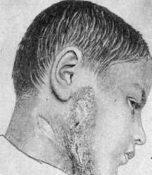
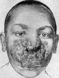
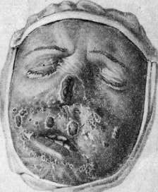
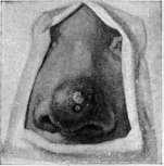
Primary affect) or inflammatory nodules with a purulent center (miliary tuberculosis of the skin). The presence in the vast majority of cases of tubercular changes in the skin characterizes an allergic restructuring of the skin tissues under the influence of the virus that has long remained in the body. Similarly, a change in the allergic state in the sense of its decrease explains the ulcerative forms of skin tuberculosis that occur in severe tuberculosis patients. Another proof of the presence of immunobiological changes are the reactions to tuberculin, which can be diverse; in most cases in patients with skin tuberculosis they are quite bright, however, there are forms in which anergy to tuberculin regularly occurs, as for example in sarcoids and lupus pernio. For the development of skin tuberculosis, the quantity and properties of the virus itself are important - massive doses lead to the development of ulcerative and pustular forms, low-pathogenic strains that have lost the property of causing follicular changes give rise to such lesions as tuberculides. Pathological-histological changes in the skin in tuberculosis are diverse. In general, two main types of reactions are distinguished: specific, the main element of which are epithelioid and giant cells, and non-specific, the main cellular element of which are lymphocytes. In some forms of skin tuberculosis, the development of a typical tubercular nodule with giant and epithelioid cells in the center and lymphocytes at the periphery predominates. Sometimes the nodules are solitary (scrofulous lupus), in other cases they merge into a common infiltrate (tuberculous lupus). Caseous decay is rarely observed and occurs in verrucous tuberculosis. In other cases, the tissue reaction consists in the formation of cellular accumulations of lymphocytes; such is often the reaction in papulo-necrotic tuberculide and some other tuberculides. A feature of tuberculous skin lesions is the presence of blood vessels in the nodules, the development around blood vessels with thickening of the walls. Changes in the epithelium are diverse - from infiltration by leukocytes, acanthosis, hyperkeratosis, development of warty growths to complete loss of epithelium in ulcerative forms. In the final stages, atrophy of the epithelium, destruction of the papillae, and development of dense connective tissue are observed. The clinical forms of tuberculosis of the skin are diverse. The most common form is tuberculous lupus (lupus vulgaris). The initial element may be a flat lupus nodule - a spot of brownish-red color, round shape, soft consistency (see separate table, fig. 4). The epidermis slightly peels off and subsequently atrophies, which is noticeable when examining the nodule by its wrinkling and fine folding. An important sign is the color of the initial lupus element - brownish, resembling the color of burnt sugar, which clearly stands out when pressed with a glass spatula (diascopy method). The consistency is determined by a pessary probe, when pressed with which a long-lasting depression remains, a sensation of pain and slight bleeding of the lupus nodule (probe test). In other cases, tuberculous lupus begins with an elevated nodule the size of a lentil, inflammatory-red in color, in the center of which a purulent crust forms. After removal of the crust, a small ulcer is seen, sitting on a not completely disintegrated nodule. Thus, lupus usually begins on the nose. Sometimes tuberculous lupus can begin with a massive infiltrate with perifocal inflammation around it (early infiltrative form of tuberculous lupus; Podvysockaya, Kaplan). Subsequently, the primary lupus elements undergo various secondary changes: peeling, crusts, ulcers or warty growths develop depending on a number of external or internal factors. Therefore, several varieties of lupus are distinguished: psoriasiform - with abundant peeling (lupus squamosus), crusty - in cases of crust formation, hypertrophic (lupus hypertrophicus) - with large infiltrate growths, ulcerative (see separate table, fig. 2) (lupus exulcerans), warty (lupus verrucosus) (see separate table, fig. 5), tumor-like (lupus tumidus) etc. (see separate table, fig. 6). The development of one or another form of lupus depends on a number of factors, among which the anatomical-physiological features of various areas of the skin are important: warty forms of lupus arise mainly on the hands, feet and fingers, psoriasiform forms - on the elbows and knees, tumor-like forms - on the earlobes etc. In addition, the routes of infection spread are important: with hematogenous infection, flat lupus usually develops, with exogenous infection - ulcerative, hypertrophic forms. With reverse development, tuberculous lupus leaves behind several discolored scar-like changes in the form of thin, atrophied, as it were, corrugated skin. Lupus nodules are often noticeable on the scars as a result of incomplete reverse development of the disease or relapse. The consequences of lupus are especially severe on the face, where it is most often located in the area of the nose and mouth. Here, partial or complete destruction of the entire cartilaginous part of the nose or tip, wings and nasal septum occurs. In the oral cavity, scarring leads to a small opening or, conversely, large defects with exposure of the gums and teeth develop. The lower eyelids are pulled down by scars, ectropion and exposure of the conjunctiva occur, sometimes the angle of the eye is pulled down or ectropion of the upper eyelids also occurs. These characteristic changes of the final stage of lupus so disfigure the face that it loses all individual features (see separate table, fig. 3). Lupus usually begins in childhood, more often after 5 years, although it can occur at any age. In addition to skin lesions, patients with lupus often have lesions of the mucous membranes of the upper respiratory tract, especially the nasal cavity. On average, 70% of lupus patients have mucous membrane lesions. In the nasal cavity, lupus is located on the cartilaginous part of the septum, the anterior ends of the inferior turbinates and in the vestibule of the nose. In the oral cavity, lupus most often affects the gums of the upper jaw in front, then the anterior part of the hard palate, the upper lip, soft palate and buccal mucosa. In the pharynx, the posterior wall and arches, the entrance to the larynx are affected. On the mucous membrane, lupus is characterized by a small-nodular, inflammatory-colored, almost always ulcerated infiltrate. The surface of the ulcer is fine-grained. In lupus patients, enlargement of lymph glands is often observed, especially on the neck. The glands are dense, the size of a forest or walnut, mobile. Bones and joints are affected in lupus patients on average in 30%. Changes in the lungs are found on average in 50%. Active exudative lung lesions are rare. Enlargement of bronchial glands, especially in childhood, can be found almost constantly. The course of lupus is chronic, with periodic improvements (in summer or with appropriate treatment) and worsening and relapses, even after apparent recovery. The lupus process is often complicated by erysipelas. On the limbs, long-existing lupus is complicated by elephantiasis, which significantly worsens its course. Skin cancers can develop on lupus scars, running significantly more severely than the cancerous neoplasm on previously healthy skin. Warty tuberculosis (lupus verrucosus) develops on the limbs in the form of a massive cyanotic-colored infiltrate with warty growths in the center, between which cracks and ulcers form. It is usually located on the fingers of the hands and feet, on the palms and soles. The disease is benign, runs very slowly and ends with a scar. It develops in adults much more often as an exogenous infection under conditions of skin damage. It can sometimes be a professional disease in people working with tuberculous material - in slaughterhouses, pathological-anatomical museums etc. In such cases, a special form of warty tuberculosis often develops - the tuberculous tubercle, differing in small size (like a pea or bean) and warty growths in the center. Hematogenous forms of warty tuberculosis are observed much more rarely, usually developing in children after measles in the form of multiple small, massively infiltrated foci with verrucous growths. The localization is diverse - on the neck, limbs, trunk. The course is significantly faster, the prognosis is unfavorable due to the presence of other tuberculous changes - in the lungs, bronchial glands and other organs. - Colliquative tuberculosis (see separate table, fig. 1) - one of the common forms of skin tuberculosis. It is characterized by the development of nodules originating from the deep layers of the skin, subcutaneous tissue or lymph glands. The nodules are the size of a forest nut, of soft consistency, the skin is initially unchanged and free, later it acquires an inflammatory-cyanotic color and fuses with the nodule. Soon in the center, a small fistulous opening opens with the release of liquid sero-purulent exudate, which dries into a crust. The course of the disease is chronic with partial scarring and further advancement of the softening infiltrate at the periphery.
The resulting scars therefore have different periods of development and are covered with bridges and papillae, characteristic of colliquative tuberculosis. Localization—in front of the ear on the cheek, along the lower jaw or the lateral part of the supraclavicular and infraclavicular area, much less frequently on the extremities. Tuberculosis colliquativa can develop as a separate node or multiple scattered nodes located in different parts of the skin. It occurs most often in childhood and much less frequently in adults. It is combined with lupus vulgaris, bone and glandular tuberculosis processes, often with phlyctenular conjunctivitis and keratitis. Lesions of the mucous membrane are encountered in colliquative tuberculosis exclusively rarely. Colliquative tuberculosis develops as a result of infection of tissues along the course, with earlier changes being tuberculosis of glands, bones, tendon sheaths, etc.; in cases of multiple lesions, it develops hematogenically. The prognosis is quite favorable, but complete recovery often occurs only after prolonged treatment. Fungous tuberculosis (tuberculosis fungosa) more often belongs to the variety of colliquative tuberculosis. Jadasson divides fungous tuberculosis into two groups that are not sharply demarcated from each other: 1) disseminated, tumor-like and 2) localized lesions. Disseminated tuberculosis tumors originate from the subcutaneous tissue, the nodes grow rapidly in depth, on the skin they open up, forming elevated mushroom-like granulations, sometimes covered with crusts and reaching considerable sizes.
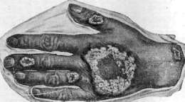
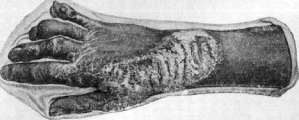
Ulcerative forms of tuberculosis of the skin (tuberculosis ulcerosa, ulcus tuberculosum). Under ulcerative forms of tuberculosis of the skin are understood those in which ulcerative decay is the main and almost sole sign. There are several clinical forms of tuberculosis ulcerosa. Miliary ulcerative tuberculosis develops most often on the mucous membrane, less frequently at the transition points of skin into mucous membrane, and rarely on the skin; its appearance is often preceded by trauma and violation of the integrity of the cover. Miliary ulcerative tuberculosis is characterized by diffuse, inflammatory infiltration, in the peripheral parts of which miliary tubercles with ulcerative central decay of greenish-yellow color are noticeable. In the center of the infiltration there is a rather deep ulcer with a granulated bottom and jagged, as if gnawed edges. The development of this form of tuberculosis is due to autoinfection with sputum or other exudate rich in tubercle bacilli. The localization of ulcerative tuberculosis is explained by the entry of infectious material onto places that can easily be infected by the patient's own excretions—the tip of the tongue, lips, skin around the anus and genital parts; less frequently it is observed on the nose, auricles and even less frequently on the skin of the trunk and extremities. Its development is facilitated by a decrease in the body's protective forces, trauma and a significant amount of the pathogen. The ulcers are painful and interfere with nutrition. The course of ulcerative tuberculosis is subacute or acute, depending on the condition of the patient. The prognosis is unfavorable.-Other ulcerative forms of tuberculosis develop on the lips, in the corner of the mouth, on the genital parts and around the anus in the form of ulcerative lesions of round outlines, with rather deep decay at the bottom, uneven bottom and slightly undermined, and sometimes even edges. These ulcers are 1-2 cm in diameter and in their form and infiltration at the base resemble the primary syphilitic lesion, which is why they received the name of tuberculous chancres. They develop as a result of autoinfection and are observed in patients with active tuberculosis. The course is subacute and rather benign. Disseminated miliary tuberculosis of the skin (tuberculosis miliaris cutis) is a manifestation of general tuberculosis with generalization of the infection. It manifests in the form of multiple scattered small red spots that quickly turn into nodules, with a pustule on top, which dry into crusts, sometimes giving the impression of small oyster shells. Tuberculides. Under tuberculides are understood such tuberculosis lesions of the skin that develop in the form of multiple scattered eruptive elements and are characterized by tuberculoid structure, rarely contain the virus, are accompanied by sharply expressed allergic reactions to tuberculin and usually develop in reverse with the disappearance of increased sensitivity. One of the typical representatives of tuberculides is papulo-necrotic tuberculide (tuberculoderma papulo-necrotica). It manifests in the form of inflammatory-colored nodules, significantly elevated above the skin. The nodule is usually the size of a lentil, round in shape; its course is as follows: in the center a pustule-like formation appears, which when opened has little liquid exudate and quickly turns into a dry necrotic plug, which does not fall off for a long time. The nodule at this time acquires a more cyanotic tint, gradually transitioning into pigmentation. The central plug falls off, leaving behind a depressed scar of round shape. The eruption of nodules is usually multiple, symmetrical; they are located in groups, most densely on the extensor surfaces of the upper and lower extremities—around the elbow, wrist, knee and ankle joints. The eruption of papulo-necrotic tuberculide occurs in bursts, and following the disappearance of the first eruption, a secondary eruption occurs, then again a fresh eruption, etc. The disease is therefore characterized by the presence of nodules in different stages of development—fresh inflammatory-colored, cyanotic nodules and scars with a pigmented border. The course of the disease is sometimes long with periodic improvements and exacerbations. The general condition is affected insignificantly, only sometimes patients complain of general weakness, weight loss and loss of appetite. Small increases in temperature may be observed. Almost a constant companion of the skin changes is enlargement of the glands in the form of a whole packet with signs of periadenitis, fused into a common conglomerate. The glands are affected more often unilaterally—on the neck, in the supra- or infraclavicular area, less frequently in the axillary fossa. Papulo-necrotic tuberculide rarely combines with other eruptions of tuberculous origin. Papulo-necrotic tuberculide can manifest in various varieties: sometimes in the form of less pronounced nodules the size of acne (acneiform tuberculide) or a lentil (papulo-necrotic tuberculide); sometimes its elements can reach the size of a hazelnut (acnitis)—a form transitional to Bazin's indurated erythema (see Bazin's indurative erythema). Scrofulous lupus of children (lichen scrofulosorum, tuberculosis cutis lichenoides) is one of the frequent tuberculides. It is expressed by the appearance of small, pinhead-sized nodules, located on the trunk rather scattered in groups. The distinguishing features of scrofulous lupus are the dirty-brown color, the small size of the nodular elements and rather abundant peeling during their reverse development. Scrofulous lupus usually accompanies other forms of skin tuberculosis—lupus vulgaris, colliquative tuberculosis, phlyctenular keratitis or conjunctivitis. In children it can also be an independent form. The general condition of the patient suffers little, however, some weight loss, grayish skin discoloration, exacerbation from other forms of skin tuberculosis, fresh eruption of phlyctenules can be noticed. The course completely depends on the general condition and usually with its improvement, reverse development of the tuberculide can be observed without special treatment. All the described tuberculides, despite the distinctive signs in clinical manifestations, have common features, namely—they represent scattered eruptions, appearing in bursts, with multiple elements, accompanied by a number of other manifestations of tuberculosis—on the skin or in the glands; tuberculides are benign forms of tuberculosis and develop in reverse with improvement of the primary tuberculosis focus and the general condition of the patient. All of them are accompanied by bright tuberculin reactions both epicutaneous and by Pirquet and intradermal, indicating a significantly expressed allergic state. Patho-anatomically, these are various forms of granuloma from an inflammatory nodule with lymphoid cells to a true tubercle.-A special position is occupied by the so-called disseminated follicular lupus of the face (lupus miliaris follicularis disseminatus faciei), which also
Fig. 1. Colliquative tuberculosis. Figure 2. Lupus vulgaris with ulceration. Fig. 3. Terminal stages of lupus; Fig. 1. Initial stages of lupus vulgaris. Figure 4. Warty tuberculosis of the skin. Figure 5. Mutilating tuberculosis of the skin. The tuberculids represent a variety of tuberculous skin diseases. The disease begins in complete health, after irritation of the skin of the face by an acute eruption of small nodules, the size of a pinhead, scattered over the skin of the face. The nodules are reddish or reddish-brown in color, elevated, sharply defined, and often have a purulent or necrotic head on top, although sometimes without signs of disintegration at the top1. Subjective sensations are absent. Other signs of active tuberculosis cannot be proven by ordinary research methods, and tuberculin reactions are often negative or weakly positive. The rash disappears as quickly as it appeared, or only gradually and slowly. The third group of tuberculous skin diseases consists of the so-called paratuberculosis. This group includes such skin lesions that are characterized by a number of common features—a special patho-anatomical structure, expressed by a reaction with the presence predominantly of lymph cells, more rarely of giant and epithelioid cellular elements. Such a lymphoid reaction occurs not only in the skin but also in other organs—lungs (marbled lungs), bones (multiple cystic osteitis), glands. The reaction to tuberculin in such diseases is negative or only weakly positive; simultaneous lesions of the usual type—lupus vulgaris, colliquative tuberculosis, or glandular lesions—are not observed in such patients. The clinical forms are: lupus pernio (see) and sarcoids (see). To this same form of tuberculous skin diseases can be attributed the ring granuloma (see Granulomas, granulomatosis, granuloma annulare), developing in the form of dense, flat, pale-colored, as if waxy, non-scaly nodules. The treatment of tuberculous skin diseases depends on a number of conditions—general manifestations of tuberculosis, the extent of the process, and its variety. Treatment must be general and local. From general measures, general strengthening agents are prescribed—cod liver oil, arsenic, iron. Specific therapy with tuberculin in its various forms is important in papulo-necrotic tuberculid, scrofulous rash, lupus vulgaris, sarcoids, lupus pernio. Diet therapy—a salt-free diet according to Sauerbruch—for lupus vulgaris gives good results. Vitamin-rich food, high in fats, and a sanatorium regime have an extraordinary effect on tuberculids and some forms of lupus vulgaris. Dry-air baths, baths with moist air of high temperature, sea bathing are beneficial factors. The main place in the therapy of tuberculous skin lesions is occupied by light therapy, and along with local irradiations, general illuminations are also of great importance. Local light therapy of lupus is used with the aim of destroying tuberculous tissue in the skin; histological studies of lupus nodules subjected to the action of ultraviolet light (Glebowsky, Delbaico, etc.) show that in them, along with changes usual for intense irradiations, there are necroses in the upper parts of the infiltrate and peculiar changes in the specific tuberculoid tissue. Ultraviolet light selectively destroys functionally weak tuberculous tissue in the dermis while sparing the normal connective tissue. Finsen initiated the light therapy of lupus, constructing for this purpose a special complex four-tube apparatus (Finsen lamp), in which the light of a carbon arc lamp passes through strong convex lenses of rock crystal, concentrating the light rays on a small space, and then through a compressor consisting of a plate of rock crystal and a plano-convex lens; the compressor, in which cold water circulates, cooling the apparatus and carrying away heat from the skin, serves to compress and anemize the illuminated area, which ensures a deeper action of ultraviolet light, since blood retards these rays. Compressors of various shapes are used depending on the illuminated area. The Finsen lamp concentrates ultraviolet light only on an area of 1 cm2, the duration of a session is 1/2-1 hour. Several hours after irradiation, an intense, relatively little painful inflammatory reaction develops, then a blister and crust form, falling off after 6-8 days; subsequently, a good cosmetically white smooth atrophic scar remains. Treatment with the Finsen lamp is carried out sequentially on small areas; limited flat lupus is suitable for this treatment. The disadvantages of treatment with the Finsen lamp—its duration and the high cost of the apparatus—cause most medical institutions to use instead the mercury-quartz lamp of Kromayer, which emits when an electric current is passed through it a light rich in ultraviolet rays from mercury vapor; as in the Finsen lamp, a compressor is used here; the duration of a session is on average 10 minutes. Other mercury-quartz lamps—the Bach lamp and the Jesionek lamp—are less suitable for initial local treatment of lupus; they find application later—after the destruction of pathological tissue (by light, surgical, or medicinal means). An excellent local agent for the treatment of lupus is natural sunlight, which has the ability, with prolonged exposure, to destroy lupous tissue just as the Finsen or Kromayer lamp does; sessions are conducted daily, their duration is gradually increased to 3 hours. For lupus of the mucous membranes of the oral cavity and upper respiratory tract, the same methods of local phototherapy are used, varied depending on the location of the process. The most accessible to solar illumination are lupus lesions of the gums, hard and soft palate, pharynx, and throat (Voznesensky). The Kromayer lamp and the Landeker lamp (Landeker; Ultrasonne), arranged like a carbon arc lamp with impregnation of the carbons with metal salts, having in its spectrum also long-wave rays, are more widely used for lupus of the mucous membranes; the Landeker lamp has a number of localizers of different sizes. Local ultraviolet light irradiations (with the Bach or Jesionek lamp) often have a favorable effect on colliquative tuberculosis of the skin (scrofuloderma), especially after preliminary surgical cleansing. General phototherapy is of primary importance in the treatment of lupus and other tuberculous skin diseases. The first place here belongs to natural solar irradiations (see Heliotherapy). Along with improvement of the general condition and blood composition, with increase of appetite and weight, skillfully conducted general light therapy has a favorable effect on allergic processes in the body, particularly in the skin, which is also proven experimentally. Lupus foci are sometimes cured by general solar irradiations alone, with isolation of the affected areas of skin from insolation (Jesionek). Combined general and local heliotherapy can at present be considered one of the best methods for treating lupus. The technique of such combined heliotherapy (Bremner) is as follows: first, for 3-4 days, a lupous area of 3-4 cm2 is subjected to very strong illumination, the duration of illumination is 2-3 hours; with good insolation this is sufficient to obtain photochemical inflammation of the third degree on the illuminated area, i.e., intense redness, blisters, and necrosis. After this, the first area is covered, and the second area nearby or far from it is illuminated in the same way. After illuminating 3-4 areas, a break is made in local irradiations and general illuminations are started (the affected areas are covered with dark material) lasting from 30 minutes to 3 hours daily, without causing intense hyperemia. After the reaction subsides, the affected area is subjected to local irradiations with small doses (for 1/2-1 hour). General irradiations with the Bach or Jesionek lamp are used with a much lesser effect; closer to the 'mixed' solar spectrum is the light of a powerful open carbon arc lamp ('arc lamp'), and indeed the light of this lamp gives a therapeutic effect close to solar; the disadvantage of treatment with the arc lamp is the excessive duration of irradiations (up to 4-5 hours), conducted in a closed room. In view of the fact that on tuberculous skin diseases and mucous membranes, a beneficial effect is apparently produced not only by chemical ultraviolet but also by thermal rays, it is recommended that when using artificial sources of ultraviolet light, patients should also be subjected to general irradiations with solux lamps (without a filter). Treatment of lupus with light is conducted either chronically intermittently or more permanently. General phototherapy is indicated for all forms of cutaneous tuberculosis (except miliary ulcerative tuberculosis of the skin) and for all so-called tuberculids.
General irradiation with sunlight or artificial light sources can only be performed after examining the condition of internal organs, since in the presence of an active tuberculous process in them, general irradiation is contraindicated to avoid exacerbation of the process. In addition, it is necessary to carefully monitor the general condition of patients, especially the temperature, throughout the entire course of general phototherapy. X-ray therapy is indicated only for hypertrophic, tumor-like, and lupus vulgaris ulcers, as well as for lupus of the mucous membranes. Dosage: 2-3 HED units with a 2-3 mm aluminum filter; the second session should not be earlier than 14 days after the first, the third (if necessary) - 1-1.5 months after the second. Often, a good effect is achieved by combined therapy with X-rays and pyrogallic ointment that destroys lupus tissue. It is rational to use X-ray therapy to 'fix' the results of surgical removal of individual lupus foci. X-ray therapy is also used in scrofuloderma, indurated erythema, and Beck's sarcoidosis. The treatment of tuberculous skin diseases with radium and mesothorium has not received widespread adoption. Method of administration and dosage of gold preparations and neosalvarsan in tuberculous skin lesions. The most commonly used gold preparations: kryzolgan (Kry-solgan) - sodium salt of complex aminoaurophenolcarbonic acid, trifal (Triphal) - sodium aurothiobenzimidazolecarboxylate, sanokryzin (Sanokryzin) - sodium aurothiosulfate glycolate, aurophos (Aurophos), etc. Gold preparations are administered intravenously; their use in ointments and solutions, locally and perifocally, has not found widespread application. Before each injection, a fresh solution is prepared in sterile water. The dosage of gold preparations varies. Dosage of kryzolgan: 0.0001-0.0005-0.001-0.005-0.01-0.025-0.05-0.075-0.1. The intervals between individual injections are set depending on tolerance, averaging from 10 to 15 days, with a total of 10-12 injections per course. The single dose can be increased only if the last injection was not accompanied by a focal reaction. In case of success, the course is repeated after 1.5-2 months. When using gold preparations, side effects are often observed, sometimes severe, which can be divided into 3 groups: 1) phenomena that occur acutely immediately or shortly after administration of the drug, similar to anaphylactic shock; 2) intoxication symptoms (elevated temperature, headache, general malaise, kidney irritation, jaundice, colitis, stomatitis, dermatitis, etc.); 3) exacerbation of the tuberculous process in internal organs. The main contraindications to the use of gold preparations: florid tuberculosis of internal organs, impaired kidney and liver function, and the island-inflammatory nature of skin rashes. Often, intravenous injections of preparations like neosalvarsan have a good effect on so-called tuberculides (papulonecrotic, Bazin's indurated erythema, etc.), especially in combination with intradermal injections of very small doses of tuberculin. The dosage and contraindications for neosalvarsan in this method of treatment are the same as for syphilis (see). Local medicinal agents are necessary for lupus vulgaris, especially for ulcerative and hypertrophic forms. Arsenical paste is prescribed (for small foci): Arsenic arsenicos. 0.6, Cinnabaris 3.0, Ung. emolliens 30.0; Pasta Plantaginis (patented preparation), 10% pyrogallic ointment, 40% resorcinol or 30-40% creosote ointments. A poultice from table salt or a 30% compress from it is a good destructive agent that also cleanses ulcerative surfaces from secondary flora. Surgical methods are varied. In the Leningrad lupus clinic, complete excision of lupus vulgaris foci and warty tuberculosis is widely used, not only for small foci but also for significant ones. Ulcerative and scab forms can be curetted with a sharp spoon followed by sprinkling with potassium permanganate, irradiation with X-rays, or application of a dry dressing. Surgical methods are the method of choice for warty tuberculosis. On mucous membranes, tuberculosis is cured by cauterizations, especially electrocoagulation (mainly lupus vulgaris and ulcerative forms of tuberculosis). Smearing with 20-25% lactic acid or trichloroacetic acid can be recommended. Care of lupus patients. Early recognition and prevention of tuberculous skin diseases. Patients suffering from advanced and disfiguring forms of lupus, usually because of the repulsive impression they produce, cannot live in a home environment, let alone in dormitories, or work, even when partial work capacity is preserved. Such patients should be placed in labor colonies at lupus clinics, where, undergoing treatment and getting accustomed to some work, they can eventually partially justify their maintenance. Totally disabled patients should, after appropriate treatment, be transferred to homes for the disabled. In the fight against skin tuberculosis, especially its dangerous form-lupus vulgaris-the organization of special medical and preventive institutions-lupus clinics-is of great importance; in the USSR, these exist in Moscow, Leningrad, Odessa, Kazan, and Sverdlovsk. Lupus clinics are institutions equipped with physical therapeutic methods, similar to hospital-sanatoriums, which are centers for the fight against skin tuberculosis of an organizational, scientific, and therapeutic nature. Prevention of tuberculous skin diseases consists primarily in carrying out basic measures to improve the socio-domestic and sanitary-hygienic living conditions of the broad masses of the population, and this in no way differs from the prevention of tuberculosis in general. Extremely important preventive significance is attached to early recognition and early treatment of tuberculous skin lesions; this is especially true of lupus. Initial cases of lupus, in contrast to its developed forms, are often relatively easy to cure. If it were always possible to recognize lupus early and carry out early treatment, we would have no severe cases at all. Lupus often begins in childhood, and in a large percentage of cases, the nasal mucosa is initially affected. Subjective sensations in this case are minimal and usually consist of nasal obstruction and the formation of crusts. Rhinoscopy may reveal a varied picture; alongside cases in which grayish-yellow and grayish-red nodules or already expressed infiltrates are visible on the mucosa, the lupus process sometimes insidiously proceeds under the guise of chronic eczema or dry rhinitis. The very earliest forms of lupus on the skin may also present certain diagnostic difficulties. In doubtful cases, histological research and others (tuberculin tests, examination of internal organs) are required for a final diagnosis. From the foregoing, it follows that one of the main conditions necessary for the early recognition of lupus is the early seeking of medical help by patients and the familiarity of a wide circle of physicians with tuberculosis of the skin and mucous membranes; the latter has particularly great importance for school physicians, pediatricians, and all rural physicians, not to mention specialists-rhinologists, dermatologists, and phthisiologists. Broad sanitary-educational work in the field of skin and mucous membrane tuberculosis, especially among the rural population, as well as among parents of schoolchildren and teaching staff, is of essential importance for earlier seeking of help. Periodic examinations of schoolchildren, mainly of the lower grades, by a rhinologist are necessary. Of no less importance are constant consultations in tuberculosis dispensaries between a dermatologist and a rhinologist. To avoid relapses, the treatment of early forms of lupus must be energetic: in such cases, surgical removal or diathermocoagulation of the affected area is preferable, followed by X-ray therapy and general phototherapy. Such patients should subsequently be under the supervision of specialists for a long time. Hygiene of the skin and upper respiratory tract, especially in childhood, also has preventive significance; for example, the habit of picking one's nose with a finger and thus chronically traumatizing the mucous membrane can be a factor that sometimes directly causes the development of lupus on the nasal mucosa in a tuberculous child; naturally, exogenous infection is also possible in this case. To carry out a planned fight against skin tuberculosis, it is necessary to maintain accurate accounting and statistics of patients with tuberculosis of the skin and mucous membranes. This accounting should be carried out by lupus clinics, and in those regions and areas where they do not exist, by those medical institutions that are engaged in the fight against skin tuberculosis-tuberculosis or venereal dispensaries or the corresponding departments of unified dispensaries. It is necessary to strive for the dispensarization of all patients with tuberculosis of the skin and mucous membranes, not only in cities but also in rural areas.
Related articles
Mentioned in
Cite this page
“Tuberculous Skin Diseases.” Soviet Medical Encyclopedia. English translation of Bolshaya Meditsinskaya Entsiklopediya, 1st ed. (Moscow, 1928–1936), ed. N. A. Semashko. https://sovietmedicalencyclopedia.pages.dev/article/tuberculous-skin-diseases/