Vegetarianism
Historical document, translated for reference. It reflects medical knowledge of the 1920s–30s and is not medical advice.
Summary
Vegetarianism is the doctrine of slaughter-free nutrition, divided historically into strict veganism ("old vegetarianism") and lacto-ovo-vegetarianism ("young vegetarianism"). This entry examines the physiological, historical, and economic aspects of vegetarian diets, noting their lower purine content, variable protein digestibility, and utility as therapeutic diets for gout, renal disease, and cardiovascular conditions.
Encyclopedia article (1928–1936)
VEGETARIANISM (from Latin vegetare—to grow), the doctrine of slaughter-free nutrition.—Forms of vegetarianism and distribution. Vegetarians consider plant food to be the only natural human food; moreover, some—"old vegetarians," or, as they are called, "raw foodists"—prohibit everything that directly or indirectly originates from animals, and some of them do not recognize cooked or fried food, eating only fruits, vegetables, and salads in raw form. "Young vegetarians," in addition to plant food, eat milk, eggs, and dairy products, which represents nutrition without the slaughter of animals. The doctrine of vegetarianism had the greatest success in those countries where, due to geographical and climatic conditions, fruits and plant foods were more accessible (subtropical countries—Italy, southern France, etc.). Leaving aside the untenable ethical side of the issue, one can approach the problem of vegetarianism from 1) historical-anthropological, 2) physiological, and 3) economic points of view.—1. Graham clarified that the structure of human teeth is very similar to the structure of the teeth of anthropoid apes; the latter feed almost exclusively on fruits, which therefore must be the natural food of humans as well.—2. Touching upon the physiological side of the issue and keeping in mind a strictly vegetarian regimen (without dairy products and eggs), it can be noted that the basal metabolism in people who have been vegetarians for many years differs very little from the basal metabolism of meat-eaters; according to the research of Benedict and Rosse, the former required 25.5 calories per 1 kg of body weight in 24 hours, and the latter 26.4 calories. Nitrogenous equilibrium can be maintained for a long time by replacing meat and milk proteins with the proteins of legumes and other plants. However, to cover the body's protein requirement, the amount of plant food must be significantly larger than that of mixed food. In addition, plant protein is closely bound to cellulose and starch, as a result of which part of the protein is lost—up to 17% (Thompson); 98% of protein is absorbed from meat, fish, eggs, and milk, whereas only 80–85% is absorbed from plant food substances, with protein absorption being even lower from certain types of plant foods: 48–75% from rye bread, 60–68% from potatoes, 60–70% from buckwheat porridge, and sometimes only about 50% from millet. The comparative weight of feces excreted per day can serve as an indicator of the poorer utilization of plant food compared to meat—with a meat diet, an average of 190 g; with a vegetarian diet, an average of 333 g (Voit). The required amount of carbohydrates, fats, salts, and vitamins for the body can be fully provided by plant food. The latter can cover the body's requirements even during heavy muscular work and sports: vegetarians can perform heavy physical labor and be good athletes. Comparative observations on muscle strength in vegetarians and meat-eaters showed that the contraction force at the beginning of work is much greater in a meat-eater than in a vegetarian, but this force drops very quickly in the former, whereas in the latter it lasts two to three times longer. Plant food is less toxic and better regulates intestinal peristalsis; it contains fewer purine bases and extractives. The disadvantages of plant food include: a) the necessity of introducing a very large volume of food to cover the body's requirements; b) plant food is utilized much worse and yields much more waste; c) it contains an insufficient amount of certain amino acids necessary for the body and does not contain hormones found in meat and certain organs of animals; d) it contains a large amount of liquid, as a consequence of which the latter is retained in tissues (Rubner, Stähelin); e) it is monotonous and requires rather complex culinary preparation. In many cases, the gastrointestinal tract adapts over time to plant food: in many vegetarians, an elongation and widening of the large intestines is noted; this was established by Hansemann, among other things, in Russian peasants who feed mainly on plant food (Russischer Darm). If milk, dairy products, and eggs are added to purely plant food (lacto-ovo-vegetarian food—slaughter-free nutrition), then all the requirements of the body, including the protein requirement, can be met very easily; from a physiological point of view, such food must be recognized as fully acceptable, and in some cases very beneficial.—3. Vegetarianism from an economic point of view. Plant food is the cheapest; not only fats and carbohydrates, but also plant proteins, taking into account their worse digestibility, are still cheaper than animal proteins. These economic conditions usually force certain groups of the population to feed exclusively on plant or lacto-plant food. In cities, however, meat nutrition costs no more than an equivalent vegetarian diet. In wealthier social groups, plant products in the food ration are usually displaced by more expensive products, particularly those of animal origin.—The doctrine of vegetarianism is an integral part of many religious and ethical systems, relying on moral motives regarding the inadmissibility of "slaughter" nutrition. Consideration of this aspect of vegetarianism goes beyond the scope of exact scientific research and presents no scientific value. Indications for prescribing a strictly vegetarian diet. A vegetarian diet is prescribed either for a short period (vegetarian days) or for a prolonged period. In the latter case, it is recommended to a) transition gradually to purely vegetarian food—with a rapid transition, many people develop severe weakness and depressed mood; b) take measures to ensure an adequate supply of protein to the body. Vegetarian days or prolonged vegetarian nutrition are prescribed for the following diseases: 1) in uric acid diathesis and gout, since vegetarian food contains far fewer purine bases and extractives (legumes containing a large amount of purine bases should be limited or completely excluded, while milk and eggs may be given); 2) in diabetes mellitus—so-called "green days" (only greens with vegetable oil are prescribed) or periodically a strictly vegetarian regimen for a more or less prolonged period; 3) in diseases of the heart and blood vessels; in case of decompensation of cardiac activity—"fruit days", instead of Karrell's milk regimen (1,000–1,500 g of fruit per day), or vegetarian nutrition for a specific period; in hypertension and severe arteriosclerosis (in these cases dairy products are also given); 4) in acute and chronic renal diseases—as a diet low in protein, purine bases, extractives, and sodium chloride; in uremia—days of nutrition with sugar, fruit juice, and fruit; 5) in obesity—supplying a large amount of greens, salads, and fruits with a low number of calories; 6) in Graves' disease—mostly lacto-ovo-vegetarian food; 7) in diseases of the nervous system, especially functional disorders, in epilepsy (limited intake of NaCl with food, enabling the accumulation of bromine in the body during treatment with its preparations), in spasmophilia in children, and tetany in adults; 8) in a whole series of gastrointestinal tract diseases; 9) in the treatment of alcoholics; plant food promotes the accumulation of fluid in tissues and, by reducing diuresis and thirst, also reduces the "thirst for alcohol."
M. Pevzner. Contents: I. Anatomy of the autonomic nervous system............480 Sympathetic system..........487 Parasympathetic system..........487 Autonomic nervous system of individual organs.........489 Anatomy of autonomic centers.......497 II. Physiology of the autonomic nervous system. Main differences between autonomic fibers and somatic ones 508 General construction of the autonomic nervous system..........509 Functions....................512 Secretory nerves of glands with external secretion 514 Secretory nerves of glands with internal secretion 522 Vasomotor nerves 524 Motor and inhibitory fibers for smooth muscles of internal organs . . . 529 Centrifugal nerves of the heart.........532 Autonomic fibers of striated muscles 539 Sympathetic innervation of receptors and the central nervous system..........548 Methods of investigation.............552 Afferent pathways and reflexes of the autonomic nervous system.....553 Physiology of autonomic centers......558 III. Autonomic reactions.............559 IV. Pathological anatomy and pathology..........560 I. Anatomy of the autonomic nervous system. The autonomic nervous system forms a part of the general nervous system and serves to innervate smooth and partly striated musculature, the heart 47B, and glands. This system consists of a central part, embedded in the central nervous system, and a peripheral part, composed of unmyelinated and myelinated nerves, which form plexuses and include a series of peripheral structures in the form of cells gathered into ganglia lying both within the walls of organs and outside them. A typical feature of the system is that the axons of the cells of its brain and spinal centers do not go directly to a specific site, but connect with the dendrites of peripheral cells, whose axons then proceed to a particular organ or to the brain, i.e., the entire peripheral autonomic nervous system is always composed of two or more neurons. The peripheral autonomic nervous system can be divided into 1) the sympathetic system, revealed by dissection and by macro- and micromethods, and 2) the parasympathetic system of conductors embedded within other peripheral nerves, distinguished on the basis of their ability to conduct stimulation, the effect of which is opposite to the action of the sympathetic system. The entire question of the autonomic nervous system is still in the stage of development. Disagreement also exists regarding the name of this system. The majority of authors accept the term "autonomic nervous system"—a name borrowed from Bichat, who used it to denote all involuntary nerves through which stimulation travels independently of our will. Gaskell's term (1886–89), "visceral system", which he contrasted with the system of the "soma" (body), based partly on morphological data, contradicts the fact of the innervation of both the "soma" and the viscera by nerves of both systems. The name "autonomous system" proposed by Langley (1908) includes the concept of the complete independence of this system from the central nervous system, which is unacceptable even to the author himself. L. R. Müller's term (1924), "vital nerves", can easily be extended to the entire motor apparatus as well. Another controversial issue is Langley's proposal to single out a third division of the system—the enteric system (the plexuses of Auerbach and Meissner embedded in the stomach and intestines); this proposal is based on the fact that this system differs in its special function and structure. A number of considerations, especially the fact that the plexuses in the stomach and intestines are entirely analogous to the plexuses of other hollow organs (urinary bladder, vagina, trachea, etc.), as well as the diversity of structural forms of cells, ganglia, and plexuses, compel us to stick to the recognition of two divisions. A third controversial point is the very possibility of isolating parasympathetic conducting pathways into an independent division. Many morphologists point out that the criterion of functional antagonism cannot be the deciding factor in separating systems. The autonomic system comprises the following divisions: I. The central autonomic apparatus, consisting of complexes of ganglion cells and fibers embedded in the spinal cord, medulla oblongata, diencephalon, and corpus striatum. II. The peripheral autonomic apparatus, consisting of the following parts: 1) the sympathetic trunk with its embedded ganglia, 2) a series of gray (unmyelinated) and white fibers extending from the trunk, 3) plexuses formed by nerves outside and inside organs, and 4) individual peripheral cells and ganglia embedded in nerve trunks and plexuses. Until recently, precise descriptions of the distribution of nerves in the stomach, intestines, heart, urinary bladder, liver, and uterus—in other words, in most of those organs whose function is essential or irreplaceable for life—have been lacking. Recently proposed dissection methods under a drop of water, as well as staining methods for the peripheral nervous system followed by clearing or transillumination of tissues, have advanced the resolution of this question, and this new methodology for investigating peripheral nerves makes it possible to freely see the nerve throughout its length up to the plexus, to see the entire course of intra-organ nerves, and also to trace nerves to their very destination and, in some cases, to distinguish the topography of axons within them. The sympathetic system originates from the ectoderm and arises simultaneously with the formation of the spinal cord. Already in the first weeks, the still undifferentiated cells of the lateral parts of the trunk segments of the neural tube from the 1st thoracic to the 2nd–3rd lumbar segments (Harman; according to Langley, to the 4th) begin to migrate out [according to Kuntz, partly from the spinal ganglia; according to E. Müller and Ingvar (1921), from the spinal ganglia]. They follow the direction of the nearest section of the anterior roots of the spinal cord (see Figure 1)—according to Kuntz, the anterior and posterior roots—and in a 4-week-old human embryo (according to Jordan and Kindred, 1926), having left the root, they position themselves externally and posteriorly to the aorta, near the developing spine, in two columns—the future sympathetic trunks (see Fig. 1a). During the same period, some cells of the columns move toward the internal organs and form peripheral ganglia. The subsequent stages consist of transformation...
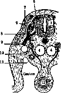
Figure 1. Diagram of the development of spinal nerves and the sympathetic trunk (Brüning, after Patterson and Cunningham): 1—spinal cord; 2—spinal ganglion (ganglion spinale); 3—white communicating ramus; 4—aorta; 5—notochord (chorda dorsalis); 6—posterior root; 7—muscle segment; 8—anterior root; 9—anterior spinal nerve; 10—ganglion of the sympathetic trunk; 11—course of the mesonephros; 12—gut.
Fig. 3. Nerve supply of the human heart. Method of D. N. Zhuravlev. Fig. 8. Nerves of the human stomach. Preparation by V. P. Vorobyov and A. A. Shamburov.
Fig. 9. Interorgan nerve trunk of the chest cavity. Preparation by A. A. Shamburov. Photomacrograph by V. P. Vorobyov.
Fig. 10. Nerves of the human gallbladder. Fig. 11. Nerve supply of the human kidney. Photomacrograph by V. P. Vorobyov.
Related articles
Mentioned in
Cite this page
“Vegetarianism.” Soviet Medical Encyclopedia. English translation of Bolshaya Meditsinskaya Entsiklopediya, 1st ed. (Moscow, 1928–1936), ed. N. A. Semashko. https://sovietmedicalencyclopedia.pages.dev/article/vegetarianism/