Feces
Historical document, translated for reference. It reflects medical knowledge of the 1920s–30s and is not medical advice.
Summary
This article from the 1928–1936 Great Medical Encyclopedia provides a comprehensive overview of the clinical examination of feces. It details the diagnostic significance of macroscopic, microscopic, chemical, and bacteriological analysis, including the influence of diet, medication, and various pathological conditions on stool characteristics.
Encyclopedia article (1928–1936)
FECES (stool, faeces, copros), the contents of the lower sections of the intestine, formed as a result of the digestive process and excreted during the act of defecation. Ancient physicians already attached great importance to the appearance of feces for the diagnosis and prognosis of disease. Leeuwenhoek was the first to subject feces to microscopic examination, but later, incorrect notions about feces led to their being completely neglected for the diagnosis of disease, and only at the beginning of the 20th century, especially thanks to the methodology and work of Schmidt and Strasburger (A. Schmidt, Strasburger), did the examination of feces receive a scientific basis and is now often absolutely necessary for making a diagnosis. The importance of examining feces is still far from being fully appreciated by practicing physicians, and it is performed by them insufficiently systematically, although for resolving the most important diagnostic questions it is comparatively simple and, not requiring especially complex laboratory equipment, can be performed even in the conditions of a district rural hospital. A systematic examination of a patient's feces consists of macroscopic, microscopic, chemical, and bacteriological analysis. This examination can be performed either with feces excreted by patients on a normal diet, or after the prescription of a so-called test diet, proposed by Schmidt for the investigation of the functional activity of the intestine, in the form of a specific standard food (see Intestine—investigation methodology). Macroscopic examination of feces considers its quantity, consistency, shape, color, odor, and the presence of food residues and pathological impurities. The quantity of excreted feces is in direct dependence not only on the quantity but also on the quality of the food consumed. Thus, according to Rubner, on an exclusively meat and egg diet, an average of 54 g of fresh feces is excreted per day, on a diet of only potatoes—about 635 g, and on only black bread—815 g; this circumstance must be kept in mind when evaluating the significance of the quantity of excreted feces, necessarily taking into account the nature of the patient's diet. In normal conditions, the quantity of excreted feces ranges from 100 to 200 g. An unusually large quantity of feces is observed in certain diseases, for example, in gastric achylia and in diseases of the pancreas (Boas). The shape and consistency of feces depend on the content of water, fat, and cellulose in them. In addition to inspection, the consistency of feces is determined by rubbing it with a wooden spatula. Normal, so-called formed feces have a sausage-like or cylindrical shape, are plastic, and are macroscopically homogeneous; such an appearance is had, for example, by feces under normal intestinal function after Schmidt's test diet. Significant deviations in one direction or another, for example, hard, mushy, and especially liquid feces, must be considered pathological. The consistency of feces depends mainly on the content of water in them; thus, in normal feces, there is 27–32% solid residue, in hard feces during constipation—40%, and in liquid and mushy feces—8–12%. Under normal conditions, with a normal diet and a good state of the intestine, 60–120 cm3 of water is excreted in the feces daily. The amount of water can increase by 2–3 times on a vegetarian regimen and fall to 15–20 cm3 during starvation. Very hard feces lose their normal shape, resulting in separate lumps (scybala) as a reflection of the prolonged stay of feces in the haustra of the intestines, or round, blackish lumps, so-called sheep or goat feces. Long flat ribbons of compressed feces are not considered typical for organic or spastic intestinal strictures; in fact, such feces are often observed with spasm of the anal sphincter and with stenoses in the lowest segments of the sigmoid flexure and in the rectum. Very watery feces are characteristic of secretory disorders of the intestine—inflammatory or nervous. Boas calls frequent feces in small portions of normally formed stool "fractional excretion" of feces. This phenomenon depends on the increased excitability of the mechanism governing defecation, thanks to which even small portions of fecal masses cause the excretion of feces (Boas). A mixture of liquid and solid feces indicates a pathological process in the intestine. Watery feces, resembling rice water, characteristic of cholera, are observed, however, in other diseases as well, for example, in giardiasis. Typical in consistency are foamy feces containing gas bubbles in fermentative dyspepsia, i.e., when fermentation processes prevail in the intestine, and in sprue. The color of feces has especially great diagnostic significance and should be determined only in relation to freshly excreted feces, because in the air, under the influence of oxygen and light, the hydrobilirubinogen of the feces turns into hydrobilirubin, which causes the feces to darken; therefore, the color of feces must be determined not only on the surface but also in the middle of the fecal masses. The color of the feces depends on the presence and state of bile pigment derivatives in them, on food and medicines, and on pathological impurities in the feces. The color of feces on a "test" diet, normally uniformly light yellow, depends on hydrobilirubin; golden-yellow, ochre-colored feces, normal for infants, indicate the presence of unchanged bilirubin and also occur (rather of a yellow-violet color) in adults with a sharp increase in intestinal peristalsis, for example, in high-seated catarrhs of the small intestine (jejunal diarrhoea). The intestinal flora has a significant influence on reduction processes in the intestine. The absence of bile excretion into the intestine gives an "acholic" stool of a grayish-white clay color (see Acholia), depending not only on the absence of bile pigments but also on a disturbance in fat absorption. In diseases of the pancreas, feces are covered with a shiny light layer of fat, the color of a melted stearin candle. The light yellow color of feces on a purely milk diet and the dark color typical of a meat diet are well known to everyone. The presence of green vegetables and fruits in food (lettuce, fresh pods, spinach, etc.) gives the feces a dark and light green coloration; chocolate, cocoa, coffee, blackberries, blueberries, black currants cause a dark coloration of the feces, from brown to blackish; the admixture of blood to food (e.g., blood sausage) gives the feces the color of tar. It is practically important to determine the change in the color of feces due to the intake of medicines. Thus, animal charcoal results in a black coloration of the feces; bismuth in small doses gives it a slate, black-gray color, iron preparations—a greenish-black, senna leaves, santonin, and rhubarb—a brown-yellow or red, calomel—green, large quantities of bismuth carbonate and barium sulfate, used in X-ray diagnostics, give light or light-gray feces, methylene blue is decolorized in the intestine and only upon keeping the feces in the air does it give them a blue or green coloration. Under the influence of an admixture of blood, feces can acquire a reddish or black coloration, and although macroscopic examination alone is not always sufficient to determine the presence of blood (see below—chemical examination of feces), it is still practically essentially important. For feces to have the color of blood, an admixture of 6% to 10% blood is necessary (Hauser). The color of feces in the presence of blood depends on the site of the hemorrhage, the speed of passage of the contents through the intestine, and the secretion of gastric juice. Under the influence of hydrochloric acid, the hemoglobin of blood shed into the stomach turns into hematin, which gives the feces a characteristic tarry appearance; in hemorrhages from lower parts of the intestine, blood is usually excreted in an unchanged form; however, even with hemorrhoidal hemorrhages (from high-seated nodes), in rare cases, tarry feces can appear due to prolonged retention of fecal masses or their reflux into the cecum (so-called retrograde transport), where the conversion of hemoglobin into hematin is also possible. An admixture of mucus and blood gives a characteristic appearance and color of meat washings to the feces in dysentery and proctosigmoiditis. The odor of feces should also be examined immediately after defecation; the usual odor of feces depends not only on skatole and indole but also on volatile fatty acids. With an abundance of meat and eggs in the diet, feces are more foul-smelling than on a milk-vegetable diet. With the prevalence of fermentative processes in the intestines, feces have an acidic odor (butyric acid); with the prevalence of putrefactive processes, they are especially foul-smelling. A disgusting, sharply putrid odor of feces indicates the presence of a decomposing tumor (or stricture) of the intestine. Feces in diseases of the pancreas sometimes have an especially characteristic odor—a sharp odor of fatty acids; a peculiar aromatic odor is sometimes encountered in the presence of ascarids (Ortner). During starvation, on the contrary, feces have no odor at all; feces that are excreted very quickly from the intestine in such acute forms of diarrhea as, for example, cholera, are also completely devoid of odor. To determine food residues and pathological components in feces, a simple inspection of the feces is not enough; it is necessary to process it accordingly; this especially applies to feces after a test diet, when it is necessary to clarify disturbances in intestinal functions and determine which components of food remained undigested.
To comminute feces and clarify the nature of residues (slags) or foreign bodies (stones, parasite particles), it has been proposed to process feces in a special sieve, passing a stream of tap water through it; Boas, Einhorn, and Strauss sieves are especially practical; one can also proceed more simply: let the feces settle in a wide (6-8-10 cm in diameter) cylinder with water and examine the particles that have floated to the surface, repeat the feces washing procedure several times, and grind the residues with water on a plate, the bottom of which is painted black with asphalt varnish. With such processing of feces, it is easy to find commonly encountered particles of food residues, mainly vegetable: peas, cabbage, beans, skins of berries and fruits, groats, and finally, sometimes small pieces of eggshells and bird feathers. Significantly more important is the determination of particles of undigested food. In some cases, for example, after gastroenterostomies or in diseases of the pancreas, macroscopically visible large quantities of undigested food are encountered: residues of meat, eggs, etc.—the so-called lientery. Significantly more often, in functional disorders of the intestine, these food residues are determined in insignificant quantities and only upon examination of feces in a ground state. This includes, first of all, residues of connective tissue in the form of white plates, most often with a decrease in the secretory activity of the stomach and the consumption of smoked meat, ham, or meat of old animals; often one can also detect residues of muscle fibers in the form of reddish or small lumps—the so-called creatorrhea. In ground feces, in the presence of a large amount of fat, it is also possible to find shiny drops of neutral fat, and even more rarely, potato residues. With this same simple technique, pathological impurities in feces are easily determined: mucus, blood, small parasites, tapeworm heads, sand, small concretions, etc. More detailed determination of these components, however, requires microscopic examination. Macroscopically, non-pathological components are determined in feces: mucus, blood, pus, and parasites. Normal feces do not contain mucus; small amounts of it, secreted by the intestine physiologically, are already digested in the intestine. Usually, mucus is easy to distinguish by the shiny, glassy appearance of the feces; often one can also see individual lumps and long strands of mucus hanging from a wooden spatula. In doubtful cases, a piece of feces is placed on a black plate or a glass slide with a small amount of water, where it is easy to see typical delicate reticular mucous formations; the addition of a few drops of Ehrlich's triacid stains the mucus a bluish-green color. Sometimes in feces, mucus is encountered in the form of peculiar large whitish-silvery strands, often confused with food residues or tapeworms; but it is enough to place them in a vessel with water to discover the finest structure in the form of delicate lace of very original patterns; this is compressed mucus, often secreted in spastic constipation and especially often in myxorrhoea intestinalis. Mucus secreted in the upper parts of the intestine is partially digested lower down; it can be detected in the form of small lumps inside the fecal masses; it is often stained golden-yellow by bilirubin and contains cell nuclei and crystals of fatty acids; a large amount of macroscopically visible mucus enveloping the feces originates from the distal parts of the intestine, and the more mucus on the surface of the feces, the lower the pathological process is usually located in the intestine. It is necessary, however, to note that contrary to the widely held opinion that mucus is always the result of an inflammatory process of the intestine, an opinion supported by the greatest specialists (A. Schmidt), it is well known that in a large number of cases it is only an intensification of the protective physiological reflex of the intestine and appears during its spastic processes under the influence of irritation of the autonomic nervous system; this circumstance has very great semiological significance. Pus is encountered in feces usually together with mucus on the surface or mixed with feces and is an expression of ulcerative processes of the lower parts of the intestine (ulcerative colitis, dysentery, neoplasm, syphilis, gonorrhea, etc.); a large amount of pus without mucus is encountered more rarely: during the emptying of abdominal abscesses (e.g., in appendicitis). Even more rarely, individual particles of tissues or neoplasms (polyps) are found in feces. Blood is encountered in feces either in macroscopically determinable quantities, pure or in combination with mucus and pus, or in the form of so-called "occult" blood, which can be detected only by chemical reactions. As already stated, bloody coloration of feces in the form of an admixture of blood or in the form of meat lumps occurs only during hemorrhages from the very lowest parts (from the rectum and sigmoid colon); usually, during hemorrhages in the upper parts of the intestine, only the color of the feces changes; they take on a characteristic tarry appearance; this does not apply to profuse hemorrhages, in which a lot of pure blood is discharged. Among other pathological impurities in feces, concretions and parasites are macroscopically determined. Of the concretions, gallstones are most often encountered—cholesterol, calcareous, pigment, or mixed. Their composition is determined by corresponding chemical reactions. Significantly more rarely, stones of pancreatic origin and coproliths are encountered. Besides the so-called intestinal sand, consisting of grains of phosphate and carbonate of lime, larger intestinal concretions are encountered—coproliths, usually consisting of food residues of plant origin impregnated with lime salts. Since barium and bismuth salts began to be used for X-ray examination, these formations, strictly speaking, false coproliths, have begun to be encountered more often. These stones should not be confused with residues of medicinal substances (pills, tablets, suppository residues). In feces, one can often detect intestinal parasites (ascarids, pinworms) or segments of tapeworms; they are easy to confuse with food residues, lumps of compressed mucus, which is why the statements of patients are always subject to verification by examining the feces for parasites and their eggs (for details, see Helminthological research methods). Microscopic examination of feces significantly supplements the results of their macroscopic analysis and is necessary in every case when it comes to the diagnosis of diseases of the digestive organs. Here, only clinically necessary microscopic examinations of feces are presented, mainly after Schmidt's test diet. For this examination, three microscopic preparations are prepared on one glass slide from feces ground with water. The first preparation (native) represents a drop of feces covered with a coverslip and studied under medium magnification of the microscope. The second preparation is prepared by adding a drop of 30% acetic acid solution to a piece of feces taken on a glass slide, followed by heating on a burner flame, and serves for the microscopic determination of fat. The third preparation is stained with a drop of Lugol's solution (Jodi puri 1.0, Kalii jodati 2.0, Aq. destil. ad 50.0) and serves to determine the degree of starch digestion and the presence of iodophilic flora. Normally, in the first preparation, one can see individual fragments of muscle fibers in the form of cylinders with rounded edges, which have partially preserved only transverse striation, individual empty potato cells, whitish or yellowish lumps of lime-soap compounds, large residues of plant food, which catch the eye first of all, and the entire field of view is filled with grains of detritus. In pathological cases, already in this preparation, it is easy to see undigested muscle fibers, most often as a result of a lack of HCl in the gastric juice or proteolytic enzymes—in the form of larger fragments of yellowish or yellow-green color with sharply defined straight edges and with preserved transverse and longitudinal striation. In addition to preserved muscle fibers, in cases of impaired gastric digestion with the absence of HCl, residues of fibrous and elastic connective tissue are also encountered; fibrous connective tissue is not always easy to recognize during ordinary microscopy; this is much more successful with the help of a polarization microscope; more compact, highly light-refracting shiny fibers of elastic tissue catch the eye. In addition to a lack of HCl in the gastric juice and insufficiency of pancreatic digestion, undigested muscle fibers, connective and elastic tissue also appear in cases of rapid passage of food through the intestine, which should be taken into account when making a diagnosis. For the functional determination of the tryptic function of the pancreas, A. Schmidt proposed microscopically determining the digestion of nuclear substance (the so-called Kernprobe); this rather complex test was subsequently simplified by Kashimodo. The patient is given a capsule containing sweetbread mixed with lycopodium powder, which has been subjected to treatment with gastric juice; such capsules are prepared by the Merck firm in Germany.
In the absence of trypsin, the nuclei of thymus gland cells can be seen under a microscope in the form of black, fused clumps lying next to easily distinguishable lycopodium. This test is positive, however, only in very severe lesions of the pancreas and the complete absence of trypsin in the feces (upon closure of the lumen of the ductus pancreaticus). In a native preparation, the presence of f a t s is also determined, occurring in the form of neutral fat, fatty acids, and soaps; they have the appearance of droplets, clumps, and needles. Droplets can be either neutral fat or fatty acids, but in a native preparation, it is difficult to differentiate them from other formations without appropriate processing; clumps can be soaps, neutral fat, or fatty acids; they are also easily confused with parasite eggs (A. Schmidt). Needles can be fatty acids or soaps; fatty acid needles are long, delicate, and sharp; fatty soaps yield shorter, coarse needles with rounded edges, gathering in clusters and clumps. For a more detailed determination of fat, the preparation is heated over a burner flame, and when it cools, a droplet of neutral fat, having melted, turns into clumps, while droplets of fatty acids yield needles, sometimes protruding from the droplet itself. It is even better to treat the preparation beforehand with a drop of a 30% solution of acetic acid, and only then pass it through the burner flame; upon cooling, it is easy to see the needles of fatty acids, which, upon repeated heating, again take on the droplet form of fat. In case of doubt, it is recommended to treat the preparation with ether, chloroform, or alcohol, whereby fatty soaps do not dissolve, while neutral fat and fatty acids do dissolve. It is even simpler to stain the preparation with Sudan III, whereby the clumps and droplets of neutral fat and fatty acids stain bright red or yellowish-red, while the soap needles remain colorless.
For the determination of carbohydrate digestion, one can practically limit oneself to staining the preparation with Lugol's solution, since in a native preparation it is not always easy to distinguish carbohydrates and decide whether a plant cell consists only of a cellulose membrane or if it contains undigested starch. This is easily determined in an iodine-treated preparation, where starch cells are stained either intensely blue, violet, or finally red, depending on the degree of digestion of the starch grains. Simultaneously, various iodophilic flora are stained blue-black in the form of Clostridium butyricum spores containing granulose, arranged in chains or clumps; rods of mature Clostridium forms, long threads of Leptothrix, and a whole series of other clumps, yeast cells, etc., are also encountered.
The rest of the native preparation is covered with the residue of food substances of plant origin, consisting predominantly of cellulose; this includes plant vessels, fruit and vegetable skins, seeds, plant hairs and villi, epidermis, etc. Precise determination of these formations is not always easy and requires special experience. To determine cellulose, the preparation is subjected to treatment with chlor-zinc-iodine (zinc chloride—30 g, potassium iodide—5 g, iodine—1 g, and water—14 cm3), whereby cellulose stains a bluish-blue color; the same is obtained when treating the preparation with iodine and 75% sulfuric acid. Besides vegetable and fruit skeleton residues, a whole series of fungal spores, various kinds of yeast, and pollen cells are encountered in the preparation, but these also difficult-to-differentiate formations do not yet have practical significance for determining pathological processes in the intestine.
Among the crystalline components of feces, one can mention crystals of cholesterol, calcium carbonate, calcium oxalate, triple phosphates, as well as crystals of medicinal substances, e.g., bismuth, sometimes salol, benzoic acid. In clumps of mucus, one can see bilirubin crystals. In feces, one sometimes also finds Charcot-Leyden crystals, which are considered by some authors (Leichtenstern, Bucklers) to be pathognomonic for the presence of parasites in the intestine; however, these crystals are often present even in such pathological processes as colica mucosa with spasms, proctitis, and chronic colitis.
Under pathological conditions, microscopic examination of feces can reveal mucus, pus, and blood in the preparation. In the presence of mucus, colorless or delicate gray threads are visible under a microscope, sometimes imbibed with yellowish bile pigments; mucus threads are more easily determined by the addition of acetic acid; iodine stains them yellow; the addition of alcohol causes the mucus threads to shrivel and become cloudy; they stain well with thionine or Methylgrün after preliminary fixation of the preparation with mercuric chloride alcohol. Often, together with mucus, one can detect p u s under a microscope in the form of a significant number of either polymorphonuclear or mononuclear elements. Sometimes eosinophilic granulocytes are found in mucus threads, which have some significance for recognizing the pathological process; they are encountered in protozoal intestinal infections (ascarids and worms), colica mucosa, eosinophilic colitis, gonorrhea of the rectum, and leukemia.
Of particular importance for diagnosis is the examination of pus in feces during dysentery, since, according to the observations of a whole series of authors, in this way one can distinguish bacillary dysentery from amoebic colitis or colitis caused by Balantidium coli. Bacillary dysentery is characterized by: a large number of pus cells, the presence of erythrocytes; pus cells are vacuolized, degenerated as a result of toxic influences; a multitude of degenerated epithelial and endothelial elements and phagocytes. Protozoal dysentery is characterized by a small number of white blood cells in the feces, the absence of plasma cells, the absence of endothelial phagocytes, and phenomena of toxic cell degeneration; white blood cells are often in a state of digestion with remnants of protoplasm at the preserved nucleus.
Microscopic examination of feces, of course, also includes the determination of bacterial flora in the preparation, and mainly intestinal parasites, both of a protozoal nature and intestinal worms, and primarily, which is practically especially important, their eggs; for the methodology of these studies, see Helminthological research methods, etc., as well as the description of various types of intestinal parasites under the microscope.
Luria. Chemical examination of feces. The average composition of feces: 8.65% nitrogen, 16.39% ether-extractable substances, and 13.82% ash. The nitrogen-containing substances found in feces consist partly of coagulated proteins of various protein formations visible even under a microscope (muscles, tendons, bacteria). Products of enzymatic protein cleavage—albumoses and peptones—are absorbed so completely during their passage through the small and large intestines that they cannot be found in normal feces. However, like amino acids, they are detected here during various diarrheas (for example, typhoid). Complex proteins—nucleoproteins and mucin—are also found in feces; the latter appears in abundant quantities in the form of mucus during catarrh. The cleavage products of nucleoproteins—purine bodies—are constant companions of nucleoproteins undergoing cleavage. Finally, diamines, as well as skatole and indole, originate from the protein particle decomposing under the action of bacteria. The amount of indole and skatole excreted per day is small and does not exceed a few centigrams. Diseases of the stomach usually have little effect on the nitrogen richness of feces. The nitrogen of feces increases slightly during obstruction of the bile ducts, and nitrogen losses are very pronounced in diseases of the pancreas. 1. The reaction of stool can be different in its various portions, on the surface, and inside the fecal masses. To determine the reaction, litmus paper moistened with distilled water is applied to stool that is as fresh as possible, freed from macroscopically noticeable impurities of blood, mucus, urine, etc. The reaction of stool is normally weakly alkaline, neutral, or weakly acidic; the pH fluctuates between 6.0 and 7.2. A significant deviation of the reaction toward acidity or alkalinity must be considered pathological. Vegetarian and fatty food gives a more alkaline reaction. Food rich in carbohydrates can cause an acidic reaction of the stool, even without causing fermentative-dyspeptic phenomena. A sharply alkaline reaction indicates increased putrefaction in the intestine and is observed in putrefactive dyspepsia. A sharply acidic reaction is obtained with insufficient digestion of fats and depends on the appearance of higher fatty acids (acidic acholic stool); this is observed especially often in fermentative dyspepsia. It is practically important to chemically determine in feces the amount and state of bile pigments, blood, fats, carbohydrates, protein, and enzymes. In addition to the usual examination of feces, the analysis of intestinal contents has recently been performed by means of the so-called intestinal capsule proposed by Reiss (v. d. Reis). A rubber tube impregnated with zinc oxide is attached to the capsule, the position of which inside the intestine is easily controlled by X-ray. The capsule is swallowed and, thanks to a valve mechanism, makes it possible to obtain intestinal contents from any segment of it for morphological, physicochemical, and bacteriological studies. 2. Determination of total nitrogen is performed by the Kjeldahl method (see Kjeldahl method). 3. Determination of bile pigments. Bile pigments in feces include bilirubin, urobilin (or hydrobilirubin, or stercobilin), and urobilinogen (or hydrobilirubinogen). a) Bilirubin. Schmidt's sublimate test. A small amount of stool is triturated with a saturated solution of sublimate, poured into a Petri dish, and left at room temperature. After a day, in the presence of bilirubin, green crumbly masses are obtained in places. b) Urobilin. 1) Schmidt's sublimate test gives a red or pink coloration in the presence of urobilin. 2) Riva-Zoja test. A small amount of feces is extracted with chloroform. Hydrochloric acid containing traces of nitric acid is added to the chloroform extract. Characteristic lines λ 550–λ 570 are obtained in the spectroscope. 3) Schlesinger's test. A small amount of feces is triturated with an equal mixture of ether and alcohol and filtered. An equal amount of a 10% solution of zinc acetate in absolute alcohol (shake before use) is added to the filtrate and filtered again. The filtrate shows green fluorescence. c) Urobilinogen. A small amount of stool is repeatedly triturated with ligroin to remove indole and skatole, which is recognized by the absence of pink coloration from the addition of an aldehyde reagent to the wash substance. The liquid is filtered, the precipitate is dissolved in alcohol, and filtered again. Ehrlich's aldehyde reagent and a few drops of strong HCl are added to the filtrate, and in the presence of urobilinogen, a red coloration is obtained. Normally, bilirubin is not found in feces; in pathological cases with rapid passage through the intestine, bilirubin can be found in the stool. Urobilin, or urobilinogen, disappears when bile ceases to enter the intestine and increases with hyperproduction of bilirubin (hemolytic jaundice). 4. Determination of occult blood. The following tests are used: a) Weber's test (see Guaiac test), b) Aloin test (see), c) Benzidine test (see), d) Gregersen's test on a glass slide (see Gregersen's test), e) Spectroscopic examination. Snapper's method. Several grams of stool are triturated in a mortar with an excess of acetone, filtered, the precipitate is washed again with acetone, squeezed out, and transferred to a clean mortar, where it is triturated with a mixture of 1 part 50% potassium hydroxide, 1 part pyridine, and 2.5 parts alcohol. 4–5 drops of ammonium sulfide are added to a few cm3 of the extract and examined spectroscopically. With a small blood content, one absorption band is visible—λ 560, with a more significant one—another between λ 523 and λ 526. 5. Determination of fats. The stool contains: a) neutral fat (glycerin ester of higher fatty acids), b) higher fatty acids (butyric, palmitic, stearic acids), and c) soaps. Normally, stool contains fat in insignificant amounts. An increase in the amount of fat occurs with insufficient secretion of the pancreas or with obstruction of the bile duct (see Acholia), or with the introduction of excessively large amounts of fat with food. In these cases, the dry residue of the stool can contain 50–80% fat. When the bile duct is closed, feces become significantly richer in fat. The average fat content in % of dry residue: normal stool—23.24%, stool with bile duct obstruction—48.65%. The cleavage of fat into glycerin and fatty acids occurs with a closed bile duct in approximately the same limits as in the norm: about 3/4 of all fat is cleaved. Fat also appears in abundance in feces in diseases of the pancreas. In these cases, fat cleavage is also reduced. Combinations of bile and pancreatic duct obstruction are distinguished by an increased content of both nitrogen and neutral fat in feces. In the ether extract of feces, besides fats and fatty acids, lecithin and cholesterol are also contained. Lecithin is always very low, and the cholesterol of bile and food is reduced by bacteria in the intestine into coprosterol and appears in feces in this form. Qualitative determination of fat has been mentioned. Quantitative determination of fat in feces: 1) A weighed amount of thoroughly mixed stool is smeared on the bottom and walls of a porcelain dish and dried on a water or sand bath at 50–60°. A small amount of dried stool is weighed, triturated with a double amount of 1% alcoholic HCl solution, and evaporated to dryness. The residue is transferred to a Soxhlet apparatus, the dish is wiped with a piece of filter paper, the paper is also placed in the apparatus, and extracted with ether for a day. Then the ether is distilled off, the residue is dried at 80° for 2–3 hours and then for half an hour at 105° and transferred to a desiccator, where the residue is dried to a constant weight. 2) Determination of neutral fat, fatty acids, and soaps according to Müller-Brugsch. A weighed amount of dried, but not treated with hydrochloric acid alcohol, stool is extracted in a Soxhlet apparatus with ether. Then the ether extract and the residue in the apparatus cartridge are examined. Volatile and higher fatty acids and neutral fat are determined in the extract; soaps remain in the cartridge. a) Volatile fatty acids. The ether extract is washed repeatedly (up to 10 times) with hot water and filtered through a smooth filter; then the acidity of the wash waters is determined by titration with n/10 alkali with phenolphthalein. The number of cm3 of alkali spent is multiplied by 0.0088, and the weight of volatile fatty acids, calculated in terms of butyric acid, is obtained. b) Higher fatty acids. After removing the volatile fatty acids, the dried extract is dissolved in a half-and-half mixture of alcohol and ether and titrated again with n/10 alkali with phenolphthalein. The amount of cm3 of alkali spent, multiplied by 0.0284, will give the weight of higher fatty acids, calculated in terms of stearic acid. c) Neutral fat is calculated by subtracting the sum of the weights of volatile and higher fatty acids from the total residue. d) Soaps. The residue in the apparatus cartridge is doused with 1% hydrochloric acid alcohol, boiled for 2 hours with a reflux condenser, then dried and extracted for 36 hours in a Soxhlet apparatus with petroleum ether.
The ether is distilled off, the residue is redissolved in alcohol and titrated with 1/10 alkali in the presence of phenolphthalein. The weight of the formed fatty acids, calculated in terms of stearic acid, is obtained by multiplying the number of cm3 of alkali used by 0.0284. 6. Determination of carbohydrates. Among the carbohydrates in feces, various types of sugar, hexoses, pentoses, and further polysaccharides, hemicellulose, cellulose, and starch are found. Fecal carbohydrates are exclusively of dietary origin. Of all carbohydrates, the determination of starch is mainly of importance. - Starch. Macroscopic and microscopic determination - see above. Chemical examination. a) Boil feces emulsified in water and filter. Upon adding a few drops of Lugol's solution to the filtrate, a blue coloration will appear in the presence of starch. b) Invert the starch into sugar by boiling the feces with 10% HCl for several minutes, filter, and perform Trommer's test with the filtrate. c) The fermentation test is based on the breakdown of starch by diastase into sugar and its subsequent fermentation under the influence of intestinal bacteria. The test is performed in a Strasburger apparatus. Approx. 5 g of feces are ground in a mortar with water, having previously determined its reaction, color, and odor, and the main vessel is filled so that no air bubbles remain in it. After filling the graduated test tube with water, the device is inverted and both test tubes are quickly put into their places. There should also be no air in the graduated test tube. The apparatus is placed in a thermostat for 24 hours. The test is considered positive if half or a third of the graduated test tube is filled with gas, the feces give an acid reaction, a lighter color, and smell of volatile fatty acids, mainly butyric acid. Insignificant gas formation can also occur during putrefactive fermentation, but the feces will then have an alkaline reaction, a dark color, and will possess a putrid odor. 7. Determination of protein. Feces contain proteins of various origins: a) dietary protein, b) bacterial protein, c) protein of intestinal secretions, d) protein of pathological intestinal discharges. For clinical purposes, the determination of protein bodies precipitated by acetic acid, serum protein, and breakdown products of protein bodies is of interest. 1) Proteins precipitated by acetic acid. a) Nucleoproteins. Normally, nucleoproteins are completely absorbed and are not found in feces. In pathological cases, increased death of cellular elements, especially in the large intestine (catarrhs, intestinal tuberculosis, proctitis, colitis membranacea, cholera nostras, typhoid fever), increases the excretion of nucleoproteins. Simon-Schlesmann test. The daily amount of feces is ground with water to a liquid consistency and left for several hours. Then it is filtered through a double folded filter or through a filter with infusorial earth. The nucleoproteins that have passed into the solution are precipitated by adding 30% acetic acid drop by drop. In an excess of acid, the precipitate easily dissolves. b) Mucin is rarely found in normal feces. It is found in pathological feces (catarrhs, typhus) containing mucus. Tests. A. Acetic acid in excess is added to the aqueous filtrate - the precipitated mucin does not dissolve in the excess acid. B. When boiled with dilute mineral acids, mucin releases reducing substances. The mucin precipitated by acetic acid is dissolved by boiling with a 7.5% HCl solution, alkalized, and the Trommer reaction is performed. c) Serum protein (serum albumin) is found only in pathological cases, and its presence indicates an inflammatory state of the intestine. Test: the aqueous extract of feces (see above) is freed from nucleoproteins by the careful addition of 30% acetic acid and filtered through a double filter. One of the usual qualitative or quantitative tests for protein is performed with the clear filtrate. 2) Breakdown products of protein bodies. These include albumoses, amino acids (leucine, tyrosine, and tryptophan). Only the determination of albumoses has clinical significance. Tests: a) the fecal filtrate is precipitated with potassium ferrocyanide and carefully heated. At 70°, the precipitate either decreases or disappears completely, and reappears upon cooling; b) albumoses precipitated by nitric acid easily dissolve in an excess of it. 8. Determination of enzymes. a) Trypsin. The material being examined must react slightly alkaline. Mett's method. A Mett tube with protein is dropped into the filtrate of feces mixed with water and placed in a thermostat for 24 hours. In the presence of trypsin, the protein column decreases from both sides. Preparation of the tube: draw filtered egg white into a thin glass tube with a diameter of 1 mm and coagulate it by boiling. Cut the tube into pieces 2 cm long, store in glycerin. More accurate, but more complex, is the Gross-Fuld method. b) Erepsin. The material being examined must have an alkaline reaction. Test: 1 cm3 of 1% peptone is added to several cm3 of fecal filtrate in a test tube. In a control test tube, the same mixture is boiled, and both test tubes are placed in a thermostat for a day. Then, the biuret test is performed with the contents of both test tubes; it will be positive in the control test tube, and negative or weakly positive in the test tube in the presence of erepsin. c) Pepsin. The material must react acid. A Mett tube with protein is inserted into the filtrate acidified with hydrochloric acid and placed in a thermostat for several hours. In the presence of pepsin, protein digestion occurs at both ends of the tube. d) Lipase. A Mett tube of the following composition is prepared: olive oil 1.0, agar powder 2.5, phenolphthalein solution 1.0, caustic potash solution 0.5, and distilled water 100.0. The olive oil is ground with agar and water to the consistency of a paste, then the remaining ingredients are added. The mixture is heated to boiling and drawn into preheated glass tubes. Then the tubes are cooled, cut, and the ends are sealed with paraffin. Test: the tube is inserted into the fecal filtrate, 1-2 drops of toluene are added, and it is placed in a thermostat for 16-22 hours. In the presence of lipase, the fatty acids formed during the breakdown of olive oil cause a color change at the ends of the tube. e) Diastase. Mett tubes of the following composition are prepared: agar powder 2.0, starch 5.0, iodine tincture 2.0, distilled water 100.0. Starch and agar are ground with a small amount of water to the consistency of a paste, and the iodine tincture and the rest of the water are added. The boiled mixture is drawn into the tubes and processed as described above. Test: the tube is placed in the fecal filtrate, which must have a slightly alkaline reaction, toluene is added, and it is placed in a thermostat for 12-24 hours. In the presence of diastase, the starch is broken down, and the blue coloration at the ends of the tube disappears. 9. Determination of indole and skatole. Indole is always present in feces; skatole is found rarely and in small quantities. An increased amount of indole and skatole is observed during increased putrefactive processes in the intestine. Indole and skatole are determined in the fecal distillate. a) Determination of indole. The daily amount of feces is mixed with water and distilled. To remove fatty acids, an excess of soda solution is added and it is distilled again. The second distillate is freed from phenolic substances by the addition of strong potassium alkali and subsequent distillation. The following tests are performed with the last distillate: 1) adding Ehrlich's aldehyde reagent to several cm3 of the distillate causes a red coloration; 2) Blumenthal's test: 0.5 cm3 of 10% vanillin solution and 1 cm3 of fuming HCl are added to several cm3 of the distillate; an orange-red coloration is obtained, which turns yellow with a few drops of 1% sodium nitrite. b) Determination of skatole. Indole is removed from the distillate by adding 10% NaOH until a slightly alkaline reaction is achieved, and then sodium β-naphthoquinone-monosulfonate is added in a small excess. After acidification, it is distilled. Skatole passes into the distillate, with which the aldehyde test is performed, giving an intense blue coloration. Bacteriological examination of feces (see Intestine - intestinal flora). The most important pathological bacteria found in feces include: the typhoid group, cholera vibrios, dysentery and tuberculosis bacteria. To isolate these bacteria, it is convenient to use the following methodology. 1) A particle of feces is taken with a platinum loop (solid feces are previously mixed in broth) and inoculated into a) two test tubes with peptone, b) two Petri dishes with Drigalski medium, and c) two dishes with Endo medium. 2) The dishes with Drigalski and Endo media are examined after a day in the thermostat. On both, colonies of Bact. coli give a red coloration, while typhoid, dysentery, paratyphoids, and Bac. faecalis alcaligenes give blue or colorless colonies. 3) Blue-colored cultures are isolated onto Drigalski medium and broth. 4) These pure cultures or suspensions in warm physiological saline are examined a) for motility; motile: typhoid, paratyphoids, Bac. Gartneri, Bac. faecalis; non-motile: dysentery; b) isolation of cultures onto sugar agar; it is decomposed by: paratyphoids, Bac.
Gartneri, Bact. coli; do not change: typhoid, dysentery, Bac. faecalis alcal.; c) litmus whey: turns red from typhoid (paratyphoids); remains blue from Bac. faecalis; d) milk: coagulates from Bact. coli; does not coagulate from typhoid, dysentery, paratyphoids, Bac. faecalis; e) mannitol-agar: blue colonies are produced only by dysentery and Bac. faecalis; f) produce indole in broth only Bact. coli. For orientation, a table is recommended (p. 788).
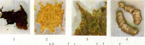
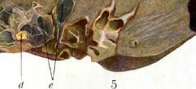
V. Blank. Feces in children. The physical and chemical characteristics of children's stool change significantly depending on various conditions: age, food, the state of the organism, etc. Normal stool. Feces of the newborn (meconium). Contents begin to appear in the intestine from the 4th month of fetal life. Feces begin to be excreted from the very first days of extrauterine life and have peculiar characteristics for the first 4-5 days. It is a viscous, homogeneous, black-green, odorless mass (Fig. 1) and is called meconium (due to its similarity to the dried juice of unripe poppy fruits). Meconium consists of secretions of the fetal intestine, epithelium, lanugo hairs, and swallowed amniotic fluid. The amount of meconium is from 60 to 90 g per day; it is excreted 1-3 times per day. From the 2nd day, the color begins to change, more brownish, then yellow masses are mixed in, and by the 4th-5th day, it is gradually replaced by the normal stool of a breastfed infant. The color of meconium depends on the presence of bilirubin and biliverdin. Chemical composition: the dry residue contains 2.5-5% nitrogen and 4.5% salts. Ash composition: insoluble in HCl-0.67%, Fe2O3-0.87%, CaO-8.0%, MgO-4.32%, P2O5-10.66%, SO3-47.05%, alkalis-24.24%. In addition to coloring substances, meconium contains bile acids (taurocholic and glycocholic), fatty acids, fat, cholesterol, and traces of blood. Indole, phenol, and stercobilin are absent. The reaction is acidic, pH is about 6. A number of enzymes are found. Upon microscopic examination, the following were found: squamous epithelial cells, "meconium corpuscles," vernix caseosa, cylindrical and goblet cells, crystals of fatty acids, bilirubin, cholesterol, neutral sodium salts, lime soaps, and mucus threads. The stool of a breastfed infant has the consistency of an ointment, is homogeneous, golden-yellow (egg yolk) in color, and has a slightly acidic aromatic odor. The color (Fig. 2) depends on unchanged bilirubin, the odor on the admixture of free butyric and acetic acids. In the first months of life, the average daily amount is about 15 g, later reaching 40-50 g, which constitutes 2-3% of the dry residue per 100 g of milk. The number of defecations in the first months of life is 3-4 per day, in the first year-2-3. When lying in the air, the stool takes on a green color, depending on the oxidation of bilirubin to biliverdin. White crumbly lumps, sometimes encountered in the feces of a normal child and previously considered to be casein, are now considered to be fatty soaps (Ca, Mg). Very often, such an ideal yellow and homogeneous stool (in a perfectly healthy child, with proper weight gain) is replaced from time to time by a stool resembling a "dyspeptic" one. The consistency becomes more liquid, an admixture of green or rapidly greening mucus appears, as well as lumps of fatty soaps. The cause of such a change has not yet been sufficiently studied. Such a stool cannot be considered pathological in a breastfed child with a good weight curve and does not require treatment. Sometimes such a stool is observed in neuropathic children. The reaction, depending on the bacterial flora, is weakly acidic to litmus, rarely weakly alkaline; the active acidity (pH) is about 5.0-6.0. The enzymes are the same as in meconium. Stool of an infant fed with cow's milk is of a denser consistency, lighter in color than when breastfed, and has an unpleasant, slightly putrid odor. The average daily amount is 40-70 g, depending on the amount of food (approximately 7.4 g per 100 cm3 of milk consumed). Frequency of feces is 1-4 times. The pale color and dryness depend on an excess of calcium phosphate. Ash composition (Michel): H 15-37% of dry residue (lime 11.68-17.69, phosphoric acid-4.28-8.31). The amount of nitrogen, according to various authors, is different: 7-30% and 3-4%. Fat is found mainly in the form of lime soaps; there is little neutral fat, and volatile fatty acids are absent. It contains hydrobilirubin, biliverdin, and indole. The stool reaction is neutral or weakly alkaline to litmus (pH-7.0-8.4), depending on the richness in salts, especially bases, and also on a greater amount of intestinal alkaline juices than with breastfeeding, which are secreted due to the high protein content in cow's milk. The appearance, reaction, and odor of stool during artificial feeding with milk formulas and with supplementary feeding depend on the properties of the food. With the addition of carbohydrates, the stool becomes darker and more acidic; when vegetables are given, it may contain residues of leaves and fibers (spinach, carrots); with the addition of meat, the color of the stool becomes pale brown with a fecal odor. Intestinal and fecal flora. The intestine of the fetus and newborn is normally free of bacteria. In individual cases, bacteria were found usually in connection with the mother's illness, e.g., cholera vibrios in cholera. Colonization of the intestine (mainly through the mouth) begins from the 4th-20th hour of life. The sparse flora of the first hours (enterococci, sarcinae, lactic acid fermentation bacteria, bacteria of Escherichia [perfringens]) is replaced from the 3rd day when breastfeeding the child by an almost pure culture of Bac. bifidus (Gram-positive flora). This change in flora depends on the reaction of the medium (pH), associated with the predominance of proteins in the newborn's intestine, and later with an increase in milk sugar. The predominance of Bac. bifidus in the feces of a breastfed child is a physiological state and is conditioned by the acidity of its stool, which stays within narrow limits of pH=5.0-5.8, not allowing the growth of other bacteria, e.g., Bact. coli and dysentery. When fed with cow's milk, Bac. bifidus is displaced by a more diverse flora, among which Bac. acidophilus and Bact. coli (Gram-negative flora) predominate. The distribution of bacteria in various sections of the intestines depends on the composition of the food and the reaction of the medium. The flora of the feces is close to the flora of the large intestine, is very abundant and diverse, and anaerobes predominate in it. Stool bacteria belong to three main types: the lactic acid fermentation group (enterococcus, bifidus, acidophilus); the butyric acid fermentation group (perfringens); and the coli-lactis aerogenes group. The feces of older children approach the stool of an adult in their physical and chemical properties and flora. Pathology of the newborn's stool. In some diseases, one can encounter discolored stool. These include: 1. Congenital atresia of the bile ducts. The stool in this case is completely white and resembles cottage cheese. In some cases, despite complete atresia of the bile ducts, the stool is colored due to the penetration of bile pigments from the blood through the intestinal glands (Fedynsky's case). 2. In congenital syphilis, the stool can also sometimes be partially discolored. Unlike these diseases, jaundice of the newborn and sepsis produce stool of normal color. Bloody stool—consisting of unchanged blood—occurs in melaena neonatorum; colored black—in sepsis. Pathology of the infant's stool. The appearance of the stool during starvation while breastfeeding is peculiar. Usually, there is constipation with the excretion of rare, dark-colored feces; this symptom is especially sharply expressed in pylorospasm. But frequent, liquid feces are often encountered, usually of an alkaline reaction due to putrefactive flora and alkaline intestinal discharge. With a sufficient supply of food, the stool becomes normal. The stool presents sharp changes during acute digestive and nutritional disorders. Simple dyspepsia. The stool is frequent up to 10-15 times, of a rather liquid consistency, often with an admixture of mucus, often green in color from the oxidation of bilirubin to biliverdin (Fig. 3). The reaction is usually acidic—pH in dyspepsia during breastfeeding is 3.2-8.4, during artificial feeding—2.8-5.4. Often, white crumbly curdy lumps, consisting of salts of fatty acids, are visible in the stool. The character of the stool and the reaction can change depending on the method of feeding: the color is sometimes whiter, sometimes darker; the consistency is sometimes more, sometimes less liquid; greenness in greater or lesser quantity; the reaction is sometimes alkaline depending on the predominance of putrefactive flora. Microscopy and chemical studies yield very diverse results and help little in the matter of diagnosis and treatment. In toxic dyspepsia (alimentary intoxication, infantile cholera), the stool can initially be the same as in simple dyspepsia; at the height of the process in severe cases, the stool becomes very frequent (20-30 times), watery, in the most severe cases resembling choleraic stool. The reaction is usually sharply acidic, less often alkaline; the color is often green, sometimes with an admixture of pus and blood. Chemically, there is a sharp increase in water, nitrogen, and alkalis in the stool. Among electrolytes, the content of Na, K, and Cl is significantly increased. A specific bacterial flora cannot be found, although some authors have attributed etiological significance to various microbes (Proteus vulgaris—Tsiklinskaya, coli-dispepsiae—Adam, Bact. coli—Medovikov, etc.). In colitis and dysentery of an infant, the stool is frequent, liquid or semi-liquid with an admixture of significant amounts of mucus, and often pus and blood. Sometimes all feces consist of masses of bloody mucus; in other cases, a significant amount of feces is mixed in, and in later periods of the disease—also pus. Sometimes the stool resembles dyspeptic stool. Bacteriologically, one can find a diverse flora; it is far from always possible, even in cases that are clinically undoubtedly dysenteric, to isolate the dysentery bacillus.
In so-called parenteral dyspepsias, i.e., disorders accompanying acute infections, among which influenza occupies a very significant place, the stool resembles dyspeptic stool, more rarely it is dysentery-like. The stool is the same in an infant with pyelitis. Nutritional disorders and chronic infection. With one-sided excessive feeding with milk and a lack of carbohydrates, a state of nutritional disorder develops, which Czerny called 'milk nutritional disorder' (Milch-nahrschaden). In this case, the so-called fatty-soapy stool is very common. It is of a lighter color, gray or almost white, dry, often excreted in the form of sausages, easily falling off the diaper (Fig. 4). Feces are rare; the odor is putrid, the reaction is usually alkaline, which depends on the predominance in these cases of processes and flora of putrefaction in the intestine. Chemically, there is a predominance of alkaline-earth soaps (Ca, Mg) over neutral fat and free fatty acids. The white color depends on the reduction of bilirubin to colorless urobilinogen. However, such stool can sometimes be observed in healthy children; it is also common when feeding with protein milk, and processes of putrefaction also predominate in the intestine, which is very important in the treatment of dyspepsia, where protein milk is indicated as a means of limiting pathologically increased fermentation processes. With excessive and one-sided feeding with flour, a flour nutritional disorder occurs, according to Czerny (Mehlnahrschaden); the stool is of a darker color, sometimes of a paste-like consistency, foamy due to an abundance of gases, foul-smelling and acidic. In chronic infections—tuberculosis, syphilis, malaria—the stool often takes on the character of dyspeptic stool (so-called parenteral dyspepsia). In intestinal tuberculosis, specific changes are observed, with mucus and blood in the stool; in syphilis—sometimes bleeding. Pathology of stool in older children. — Acute diseases of the gastrointestinal tract. 1) Acute gastroenteritis (alimentary, associated with gross violations of diet, and infectious). The stool is liquid, abundant, watery, with gases and often with a strong odor; sometimes—an admixture of mucus and blood. 2) In cases of poisoning, the stool can be very diverse depending on the nature of the poison. In children, poisoning with mushrooms (fly agaric, death cap), berries (wolfberries), plants (henbane, thorn apple), roots of poisonous plants, sausage and fish poisons are more common. In these diseases, the stool can be as in acute gastroenteritis or as in colitis. Often in feces, one can find residues of undigested fiber particles, berries, and grains. The stool can be dysentery-like, with mucus and blood, in cases of poisoning with heavy metals, acids, and alkalis. 3) Acute infections. Changes in the stool are sharp and very characteristic in infections affecting the intestinal tract: cholera, typhoid fever, and dysentery. In cholera, the stool can be completely typical, as in an adult, and appear in the form of so-called 'rice water,' consisting of water with floating masses of mucus. The stool is usually discolored, the reaction is alkaline. Sometimes there is an admixture of blood. Upon bacteriological examination—cholera vibrio. But in some cases, the stool can be very atypical, of liquid and semi-liquid consistency with an admixture of mucus. In typhoid fever and paratyphoid in children, the stool does not always have the typical appearance of 'pea soup,' constipation with scanty, solid feces is often observed. In case of bleeding—an admixture of bright red blood. From the feces, one can isolate the typhoid bacillus, however, for diagnosis, the method of plating the bacillus from blood onto bile (first 10 days) and the Widal reaction in later periods of the disease are more accepted. In bacillary dysentery, the stool can be very diverse.

[Garbled text]
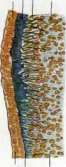

[Garbled text]
Figure 2. Normal stool during breastfeeding. Figure 3. Dyspeptic stool. Figure 4. Fatty-soapy stool. Figure 5. Hemorrhagic infarcts of the lung... [The remainder of this paragraph consists of garbled text and figure captions for unrelated medical illustrations].
Feces depending on the form and period of the disease. In mild cases, the stool may remain fecal, semi-liquid with an admixture of mucus and green. In more severe cases, at the height of the disease, the stool consists of a mucous-bloody liquid. In the most severe cases, the feces consist of scanty bloody (coffee grounds) discharges. In later periods—an admixture of pus. Upon bacteriological examination, it is possible in fresh cases in the first days of the disease to isolate the dysentery bacillus; however, the percentage of such findings is still small (40-70%). In amoebic dysentery, the stool is very similar to that in bacillary dysentery; the difference lies in the fact that the mucus is stained with blood and represents a mass resembling 'raspberry jelly,' in contrast to the unstained mucus of ordinary dysentery. Upon microscopic examination, amoebae are found. Other acute infections, although they do not directly affect the intestinal tract like those described above, can in a number of cases cause intestinal function disorders and peculiar changes in the stool. Infections that often cause disorders include influenza and measles. With them, the stool, especially in young children, becomes very liquid, dyspeptic, with an admixture of mucus, and sometimes blood. Very sharp changes in the stool are observed sometimes, especially in severe cases, in relapsing fever: mucus, blood appear, and the stool resembles dysentery. In a number of other infections (for example, scarlet fever, smallpox, lobar pneumonia), in very severe cases, with phenomena of significant toxemia, a liquid green diarrhea may be observed at the beginning of the disease. Sepsis, complicating various infections, is also often accompanied by such diarrhea. Some infections do not cause changes in the stool at all. Changes in the stool are very characteristic in infectious jaundice (Botkin-Weil disease). The stool is sometimes completely or partially discolored, clay-like in appearance. Chronic diseases of the gastrointestinal tract can cause very diverse stool disorders, with a predominance of either putrefaction processes or fermentation processes depending on the causes that caused them (fatty diarrhea, fermentative dyspepsia). The appearance of feces in so-called colitis membranacea is peculiar—fecal, often completely formed masses are covered with mucus in the form of ribbons and strips. Similar to this are feces in so-called mucous colitis, the genesis of which is associated with overfeeding with proteins and in which a large amount of mucous masses in the form of membranes and shells is also observed. Severe intestinal disorders were seen during starvation. The stool took on the character of acute colitis. Chronic infections. In intestinal tuberculosis, diarrhea develops, often with mucus and blood and with Koch's bacilli in the feces. A tubercular process, even not localized in the intestine, especially in young children, can cause prolonged disorders. In tubercular meningitis, constipation is common. Malaria usually does not affect the stool, and only in severe cases can dysentery-like feces be observed. Congenital syphilis in a young child can cause prolonged diarrhea. An admixture of pus in feces is observed in a number of diseases: in the late periods of typhoid and dysentery, relapsing fever, cholera, in abscesses that have broken into the intestine, ulcerative processes of the intestine. Blood in the stool can be observed in hemorrhagic diathesis (purpura), ulcers of the intestine and stomach, nephritis, intussusception, sepsis, syphilis, relapsing and typhoid fevers, dysenteries, poisonings, blood diseases (leukemias), hemorrhoids. Significance of feces examination. Chemical examinations of stool in children for clinical diagnosis are currently given little importance. In infancy, stains for fat, fatty acids, and starch are used. In older children, if necessary, standard examinations for the presence of bile pigments, indican, and blood are performed. Examinations for helminth eggs are of great importance. In some cases, bacterioscopic and bacteriological examinations (tuberculosis, typhoid, dysentery) are used, as well as examinations for enzymes. A. Koltynin.
Related articles
Cite this page
“Feces.” Soviet Medical Encyclopedia. English translation of Bolshaya Meditsinskaya Entsiklopediya, 1st ed. (Moscow, 1928–1936), ed. N. A. Semashko. https://sovietmedicalencyclopedia.pages.dev/article/feces/