Roseola
Historical document, translated for reference. It reflects medical knowledge of the 1920s–30s and is not medical advice.
Summary
This article from the 1928–1936 Soviet medical encyclopedia defines roseola as small, bright red macules resembling lentils, which are manifestations of various skin diseases. It details four groups of roseola: febrile (e.g., measles, rubella), infectious (e.g., syphilis, typhus), drug-induced, and emotional. The text provides a detailed clinical description of syphilitic roseola, including its histology, diagnosis, and recurrence patterns.
Encyclopedia article (1928–1936)
ROSEOLA (roseola), small hyperemic bright red spots the size of a lentil, representing only manifestations on the skin of various diseases. Thus, according to Daria, four groups of roseola are distinguished: 1) febrile roseolar rashes (measles, rubella), 2) infectious symptomatic roseola (in syphilis, typhus, cholera, smallpox, spinal meningitis and some forms of septicemia), 3) drug-induced roseola (see Dermatitis), 4) emotional roseola (in excitement) in the form of rapidly disappearing spotted or reticular redness appearing in some individuals on the chest, neck, and shoulders upon undressing, which is a physiological phenomenon similar to the redness of the face upon excitement. Syphilitic roseola (spotted, or erythematous syphilid) consists of round or oval sharply delimited pale red or bright red spots lying at the level of the skin or slightly elevated above it due to hyperemic prominence, which never peel and do not itch. With the first eruption, usually an abundant symmetrically located fine-spotted roseola occurs, reaching a size of about 1/2 cm in diameter. The favorite localization is the lateral surfaces of the trunk, the skin of the abdomen, the flexor surfaces of the upper extremities, less often the neck and face. In emaciated and weak people, especially in women, upon the appearance of roseola, a slight fever, headaches, loss of appetite, and general malaise occur. Under the influence of specific treatment or without it, roseola gradually pales and disappears without a trace. Roseola develops on the 4th–6th week after the appearance of the chancre. Its duration of existence is from 2 to 6 weeks, which depends to a large extent on whether the patient underwent specific treatment. In late recurrences, roseola appears mostly scanty and pale, asymmetrically scattered in the form of larger spots with a tendency to group in the form of arcs and ring-shaped figures. Such ring-shaped or garland-like large-spotted roseola, sometimes recurring up to 5 times, may appear even after 3–4 years after infection (late recurrent roseola, roseole tardive Fournier).- Histologically: hyperemia and slight small-cell infiltration of the papillary and subpapillary layers of the skin around dilated vessels, the walls of which are thickened. Pale spirochetes are found mostly in scanty quantity.- Recognition is usually easy, but in doubtful cases it is necessary to look for accompanying signs of infection (residues of sclerosis, swelling of lymph nodes, lesions of mucous membranes, focal hair loss, etc.) or perform the Wassermann reaction. In the differential diagnostic respect, one has to take into consideration Gibert's pityriasis rosea, exudative spotted erythema, ringworm, marbling, blue spots from flea bites.- Roseola in febrile diseases (measles, rubella) is characterized by a pale-pink color, the spots quickly acquire a saturated redness, merge and turn into a papule; the eruption is accompanied by a sharp rise in temperature; the regression of the measles rash is accompanied by peeling; roseola in rubella usually passes without peeling and pigmentation. Drug-induced roseola (after taking balsamic preparations) is characterized by itching, bright red color.
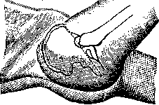
ROSER-NEELATON LINE (Roser, Nelaton), a line drawn between two points: 1) spine of the anterior superior iliac spine, 2) tuber ischii. Normally, the greater trochanter of the femur (trochanter major) is located on this line. In cases of displacement of the femoral head (dislocation, destruction of the head by an inflammatory process) or shortening of the femoral neck (fracture), the greater trochanter is located above (rarely below) this line. The examination is best performed in the position of the patient on the side with the thigh brought to the abdomen, connecting both points (sp. oss. il. sup. ant. and tub. ischii) with a thin thread. The Roser-Neelaton line in the clinic of diseases of the hip joint and femoral neck serves as a great aid, allowing one to make the correct diagnosis without X-ray examination.

Related articles
Mentioned in
Cite this page
“Roseola.” Soviet Medical Encyclopedia. English translation of Bolshaya Meditsinskaya Entsiklopediya, 1st ed. (Moscow, 1928–1936), ed. N. A. Semashko. https://sovietmedicalencyclopedia.pages.dev/article/roseola/