Sideroscope
Historical document, translated for reference. It reflects medical knowledge of the 1920s–30s and is not medical advice.
Summary
A sideroscope is a device based on the properties of a magnetic needle to deflect in the presence of even the smallest particles of ferromagnetic metals. It is used in ophthalmology to detect and locate iron or steel fragments in the eye. The instrument was first proposed by Asmus in 1894 and later improved by Hirschberg.
Encyclopedia article (1928–1936)
SIDEROSCOPE (from Greek sicleros-iron and scopeo-I look), a device based on the properties of a magnetic needle to deflect in the presence of even the smallest particles of ferromagnetic metals. A sideroscope is sometimes used in ophthalmic practice to determine the presence and localization of iron or steel fragments in the eye. The original model of the S. was proposed by Asmus in 1894 and was improved by Hirschberg (Fig. 1 and 2), who introduced changes that significantly simplify the use of the device. The main part of the latter is an astatic magnetic needle freely suspended on a silk fiber inside a glass cylinder by means of three screw legs. The device is set in such a position that the needle, having a strictly vertical direction, does not touch the walls of the cylinder. To capture the most insignificant oscillations of the needle, a small flat mirror (Gauss) is attached to its middle part, to which light is directed from a lamp. The beam of light reflected from the mirror is cast onto a screen with a special scale placed near the apparatus. When the eye is brought to the device in the presence of iron or steel, the needle comes into motion, which is communicated to the mirror, and thanks to the movements of the latter, the beam of light reflected from it moves on the screen along the scale. The amplitude of the needle's oscillations allows one to judge to a certain extent in which area of the eye the foreign body is located. In general, the device is characterized by great sensitivity, and with its aid it is possible to determine in the eye fragments weighing less than 0.001 g. However, the S. also has significant drawbacks, since the slightest shaking of the wall on which it is mounted causes the needle of the device to move, which hinders the examination, especially with small sizes of the foreign body. The same is observed under the influence of iron or steel objects found accidentally nearby, and especially under the action of the magnetic field around the wiring of the lighting network or tram wires, etc. Due to these drawbacks, the S. is relatively rarely used for the diagnosis of foreign bodies, yielding to the strong electromagnet and the X-ray.

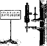
Fig.
Sideroscope according to Hirschberg.
a. crushing. SILOSIS, sycosis, sycosis vulgaris, s. simplex, s. staphylogenes, s. non parasitaria (from Greek sy-con-grape), a chronic superficial follicular staphylococcal dermatitis observed almost exclusively in men on hairy parts of the face, less often in the axillary regions, on the pubis, and on the head (see separate table, Fig. 1). The so-called parasitic sycosis is a deep trichophytosis, sometimes similar to ordinary sycosis due to its localization and clinical picture. The etiology of sycosis is complex and not yet fully elucidated. The microbial flora of the process is the golden, white, staphylococcus, indistinguishable by modern methods of investigation from ordinary staphylococci. However, there are no data proving the contagiousness of sycosis as such or explaining the chronic course and tendency to recurrence by special properties of the pyococci. It is therefore necessary to think that it is precisely in the organism itself that there are some prerequisites determining the disease of sycosis. Cases of generalized sycosis with involvement of the face, axillary regions, pubis, etc., indicate a systemic involvement of the follicular apparatus. It is possible that various factors (nervous, endocrine, etc.) create corresponding biochemical prerequisites in the follicles for the multiplication and stable existence of pyococci. The social significance of sycosis is determined by its frequency and its role as a factor of temporary disability. Among 1,044 admissions of male patients with various forms of pyoderma in 1928-32 at the Tarvonsky Hospital in Leningrad, sycosis patients accounted for 29.1%, for whom 9,634 working days were lost, and the average length of stay of patients who were hospitalized once was 30.5 days. From the pathological-anatomical side, sycosis is characterized by the development of an intracapsular pustule occupying the hair follicle funnel and rarely deeper. The walls of the follicle are never destroyed, but can be penetrated by a significant number of segmented leukocytes. Perifollicular infiltration usually has a round-cell character. In old cases, signs of chronic inflammation are found in all layers of the skin. The pathogenesis of sycosis is reduced mainly to infection of hair follicles by pyogenic staphylococcus, and the duration of the course and recurrences are explained either by the absence of development of immunity inside the follicles or by its periodic fluctuations. Along with the recognition of exogenous infection of follicles by pyococci, one cannot deny the possibility of mutation of the always present in the follicles non-pathogenic staphylococci into pyogenic forms. Various biochemical intracapsular processes, dependent on nervous and endocrine and other moments, as well as external influences traumatizing the follicle openings, can contribute to this assumed mutation. Acute recurrences occurring simultaneously over a large surface and in many localizations speak in favor of such a possibility. The influence of profession (lubricating oils, dust, etc.) is undeniable, but it is not decisive and affects rather people predisposed to the disease. In this sense, the role of chronic rhinitis preceding facial eczema, etc., can also be understood. Course and symptoms. Depending on localization, spread, and outcome of sycosis, several clinical types are distinguished, connected by transitional forms. The most common clinical form is sycosis of the upper lip. Chronic rhinitis is often found in it. The course is chronic, usually recurrent in separate flare-ups, between which there may be more or less long remissions. In the acute stage, superficial follicular pustules predominate, penetrated in the center by a hair, sitting on the background of inflamed hyperemic skin. The contents of the opening pustules dry up in crusts, merging with each other, and inflammatory phenomena between follicles can intensify to the degree of oozing, simulating the picture of eczema (eczematized sycosis). A combination with signs of seborrheic eczema is also possible, especially in the eyebrow area (sycosis-form eczema). In the subacute stage, infiltrative phenomena predominate: the number of pustules can be very insignificant, but there is a scaly desquamation on the background of reddened and thickened skin. In other cases, deeper dense furunculoid infiltrates are formed or more superficial soft elastic nodules, standing out above the level of the skin. On the hairy part of the head, sycosis resembles impetigo, differing from it by the duration of the course and persistence of recurrences. The so-called dermatitis papillaris capillitii Kaposi is apparently a variety of sycosis and is characterized by the formation on the back of the neck and the adjacent part of the hairy part of the head of dense nodules and plaques of pale or violet-red color. Generalized sycosis is characterized by involvement not only of the face, axillary regions, and pubis, but also of the lower extremities. Lymphadenitis in sycosis is observed very rarely. The duration of the disease and its persistence in relation to therapy can affect the psyche of the patient and become the cause of a neurasthenic-hypochondriac symptom complex. Diagnosis is based on characteristic morphological signs and the course of the disease. It is important to distinguish ordinary sycosis from the so-called parasitic sycosis. The latter is characterized by asymmetrically located, sharply delimited, rounded, standing out above the level of the skin plaques of bright red color. On palpation they are dense, and when pressed from the sides, abundant drops of pus are squeezed out from the follicle openings. The scales on the surface of the plaque are usually significantly thinned, break off, or even fall out completely. The causative agent is as a rule a fungus of animal origin. Sycosis differs from eczema by the predominance in the clinical picture of follicular inflammatory nodules and pustules. It differs from impetigo by this, as well as by the duration of the course. It should be borne in mind, however, that eczema and impetigo of the hairy parts of the face can give impetus to the development of sycosis. The prognosis of sycosis regarding complete recovery is doubtful. However, it is possible almost always to achieve a cosmetic effect and maintain this effect. Thus, from the point of view of work capacity, the prognosis is favorable, although one should always keep in mind the possibility of recurrence. Treatment of sycosis does not yet possess methods guaranteeing complete cure and prevention of recurrences. Therefore, the goal of therapy is the fastest possible cosmetic effect and the earlier restoration of work capacity. Specific therapy (auto- and polyvaccination) has only auxiliary significance. Bezredka's filtrates do not give special results. Among physiotherapeutic procedures, roentgenotherapy has significance, applied either for the purpose of epilation or in fractional doses, as in the treatment of eczema. Epilation of affected areas (roentgen or manual) gives in combination with sequential local disinfecting therapy the best results. For the latter, especially dyes (gentian or methyl violet or diamond green), applied in the form of ointments (1%), aqueous lotions (1:1000) and 70° alcoholic solutions for lubrication (1%) can be recommended. Good results are also given by electrophoresis with an aqueous solution of the dye. Great significance also has sulfur preparations in the form of ointments or dustings. Tar preparations are also applied. In acute cases, lotions (Burov's liquid, aqueous solution of dye, etc.) are expedient. To prevent recurrences, frequent shaving and daily wiping of affected areas with 70° alcohol for a long period of time are recommended. Existing rhinitis must be subjected to thorough therapy. Work in dusty and metalworking professions associated with the use of lubricating oils is contraindicated in sycosis, and therefore it is expedient to transfer patients with persistent recurrences to other work.
S. Rosenthal.
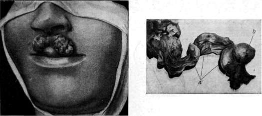
Figure 1, Sycosis parasitaria.
Figure B. Aneurysm (b) of the splenic artery (a).
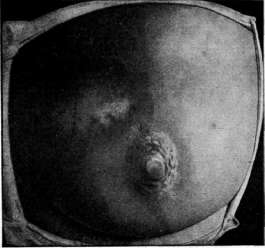
Figure 3, Scleroderma in plaques.
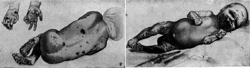
Figure 4. Congenital syphilis.
Figure B. Congenital syphilis.
Related articles
Cite this page
“Sideroscope.” Soviet Medical Encyclopedia. English translation of Bolshaya Meditsinskaya Entsiklopediya, 1st ed. (Moscow, 1928–1936), ed. N. A. Semashko. https://sovietmedicalencyclopedia.pages.dev/article/sideroscope/