Xeroderma Pigmentosum
Historical document, translated for reference. It reflects medical knowledge of the 1920s–30s and is not medical advice.
Summary
Xeroderma Pigmentosum is a rare genetic disorder characterized by dryness and pigmentation of the skin, progressing to atrophy, telangiectasia, and malignant transformation. The disease begins in early childhood, is inherited as an autosomal recessive trait, and is associated with heightened sensitivity to ultraviolet radiation.
Encyclopedia article (1928–1936)
XERODERMA PIGMENTOSUM (from Greek xeros-dry and derma-skin) (synonyms: melanosis lenticularis progressiva, epitheliomatosis pigmentosa, liodermia essentialis cum melanose et teleangiectasia, atrophoderma pigmentosum et atrophic, parched skin), a disease first described with almost exhaustive completeness in 1870 by Kaposi. X. pigmentosum occurs rarely. According to the calculations of Siemens and Kohn from 1870 to 1925, only 333 cases in 222 families have been described in the world literature, of which 146 cases are single in the family and 187 are multiple (in 76 families). Men and women get sick equally often. The characteristic signs of the disease are dryness and pigmentation of the skin. Subsequently, scaling, atrophy, scar-like changes, telangiectases, cracks, ulcers, eczematizations [see separate table (Vol. XIV, art. 231-232), Fig. 7], blepharitis with loss of eyelashes and subsequent formation of keratitis, atresia buccalis, and finally epithelial formations develop, which usually undergo malignant cancerous degeneration. Cases have been described when connective tissue neoplasms of the type of angiomas, sarcomas, etc. developed. The disease usually begins in the first three years of life (80% of all cases), more rarely in adolescence and extremely rarely in mature age (Terebinsky's case - onset at 31 years, Favre's case - at 64 years). The first signs of the disease are often detected in the spring and summer months, and in some cases, the development of typical X. p. is preceded by dermatitis of the solar erythema type. The course of the disease is always chronic, steadily progressive, with periods of improvement and lull. According to Siemens, two-thirds of all patients die before the age of 15. However, cases have been described where the pathological process in the 20-30-year age group paused and patients lived to old age (Sh.'s case - up to 60 years, Matzenauer's case - up to 66 years, and the Herxheimer-Hildebrand case - up to 70 years).
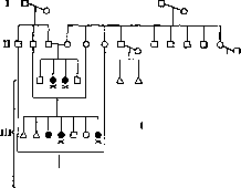
Histological research: chronic inflammation with outcome in atrophy, abundant pigment deposition, neoplasm of blood vessels, breakdown of elastic and connective tissue fibers, and atypical proliferation of epithelium in the form of cancerous tumors. - Etiology and pathogenesis. X. p. is a genotypic disease, inherited as an autosomal recessive trait. According to Siemens, in 17% of all described cases, consanguinity is noted. In cases where genealogy was carefully studied, this percentage rose to 59. Consanguinity was noted in the case that Olesov had to observe in the clinic of skin diseases (Fig. 1). An interesting case is described by Velhagen: three healthy brothers married three healthy sisters; in two of these marriages, some of the children turned out to be sick (Fig. 2). - X. p. is clinically manifested only in homozygotes, i.e., only in the case when the predisposition to this disease is inherited simultaneously from both mother and father; in those cases where predisposition is received only from one parent (in heterozygotes), clinical manifestations of the disease are not noted. - The usual onset of X. p. disease after prolonged exposure to the open air and especially after the first insolation is explained by the increased sensitivity of the skin to ultraviolet rays of any wavelength, and their action on the skin is slow and deeper; the altered sensitivity of the skin in X. p. is noted to X-rays and alpha rays, and normal - to the solar spectrum and chemical irritations (Martenstein). The blood does not contain photodynamic substances (hematoporphyrin, etc.), and its injection into the vein of animals does not sensitize them. Thus, ultraviolet rays as an external factor are the manifestation of the gene causing X. p.
Diagnosis of the disease in the presence of clinical manifestations does not present difficulties. Differentiation must be made with freckles, scleroderma and sometimes with leprosy. - Treatment is symptomatic - it aims to protect the skin from the action of sun rays, for which hats, gloves and protective skin ointments and pastes containing quinine and tannin are recommended. Tumors are removed surgically, by radium, X-rays and electrolysis.
Related articles
Mentioned in
Cite this page
“Xeroderma Pigmentosum.” Soviet Medical Encyclopedia. English translation of Bolshaya Meditsinskaya Entsiklopediya, 1st ed. (Moscow, 1928–1936), ed. N. A. Semashko. https://sovietmedicalencyclopedia.pages.dev/article/xeroderma-pigmentosum/