Biological Analysis
Historical document, translated for reference. It reflects medical knowledge of the 1920s–30s and is not medical advice.
Summary
Biological analysis is a branch of applied hydrobiology used in sanitary practice to determine the degree of water pollution. It relies on the study of biocenoses—complexes of organisms that develop in specific environmental conditions—to serve as indicators of water quality.
Encyclopedia article (1928–1936)
BIOLOGICAL ANALYSIS, a branch of applied hydrobiology that allows for the determination of the degree of water pollution and is currently finding wide application in the field of sanitary practice. Biological analysis proceeds from the following tenet of modern biology: the development of both individual organisms and the complex complexes (biocenoses) formed by them in nature is in direct
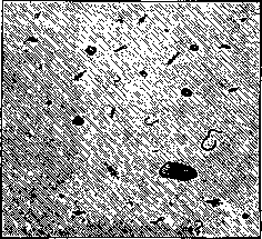
Figure 1. Production of the bottom of the Moscow River in an unpolluted area. The figure shows organisms developing on 1/10 sq. m of the bottom surface. The background corresponds to the shade of the soil (sand).
dependence on the external conditions of their surrounding environment. The presence of certain conditions determines the development of some organisms, while simultaneously suppressing the development of others, and leads to the formation of specific complexes—biocenoses—corresponding to these conditions. The study of the flora and fauna of polluted waters has shown that, depending on the degree of water pollution by organic substances, different conditions are created in it, promoting the development of some organisms and suppressing the development of others. In the most polluted waters, certain organisms are found; in less dirty ones, others; and in clean ones, still others. Each degree of pollution corresponds to its own characteristic complex of aquatic organisms, its own biocenosis. Figures 1 and 2 clearly depict the results of a thorough analysis and count of two soil samples taken from different places along the course of the Moscow River. Figure 1 corresponds to a sample from a completely clean area, above the city of Moscow, and Figure 2 to a sample from a heavily polluted area within the city of Moscow. Each sample covered an area of 0.1 sq. m. All larger (macroscopic) organisms found in the samples are fully depicted in the diagrams. The difference in both qualitative and quantitative terms is striking. In the clean soil (see Figure 1), there is a fauna that is rather weakly developed quantitatively, but represented by rather diverse forms: here are various mollusks, insect larvae, and bristle worms. None of these organisms are found in the polluted soil. Here, one form of bristle worm is developed in enormous abundance, and it is a form that is not present in Figure 1. Figure 3 (diagram) also very clearly shows how strongly the pollution of river water by paper mill effluents influences the change in the aquatic organisms inhabiting the water. Forms that developed in the river before pollution disappear from it completely after pollution and again

Figure 2. Production of the bottom of the Moscow River in a polluted area. The figure shows organisms developing on 1/10 sq. m of the bottom surface. The background corresponds to the shade of the soil (silt).
begin to appear only then (after 15–20 km), when the river water, according to the chemical data presented on the same diagram, is already significantly purified through the processes of natural self-purification. Along this same polluted stretch, in place of the disappeared forms, others appear that were not previously encountered in the river, adapted to life in heavily polluted waters. However, the above-mentioned tenet of biology can also be applied in reverse. If a certain complex of conditions leads to the development of certain organisms and biocenoses, then, obviously, conversely, by the presence of certain organisms or by a known biocenosis observed in reality, one can draw a conclusion about the conditions that caused their development. If organisms are discovered in water that can develop only in heavily polluted waters, one can conclude that the given water has a high degree of pollution. For this conclusion to be sufficiently firmly grounded, one only needs to be sure that the found organism (or organisms) is indeed encountered only in heavily polluted waters and can never be encountered in cleaner waters. One must be sure that it can indeed serve as a "representative" organism, an "indicator" organism. It turned out that of all the representatives of freshwater flora and fauna, only a few can serve as such living reagents for organic pollution. The creators of modern, scientifically grounded biological analysis—the German scientists Kolkwitz, a botanist, now a professor at the University of Berlin, and Marsson, a zoologist (died in 1908)—identified about a thousand such indicator organisms, uniting them under the general name of saprobic (putrefactive) organisms into a system of saprobity. Practically, biological analysis arose from the microscopic analysis of sediments, which often accompanied chemical and bacteriological studies of water. In this, the presence in the water of such impurities as specks of bluing, fragments of digested striated muscle fibers, starch grains, cotton and linen fibers, etc., was noted first and foremost, the presence of which can often give very important indications of the origin of pollution in the water. Schematic characterization of saprobic zones. [Table omitted for brevity in translation, but the content describes chemical composition, oxygen conditions, biochemical character, iron compounds, putrefaction tests, oxygen sources, bacterial content, and organism groups across polysaprobic, alpha-mesosaprobic, beta-mesosaprobic, and oligosaprobic zones.]
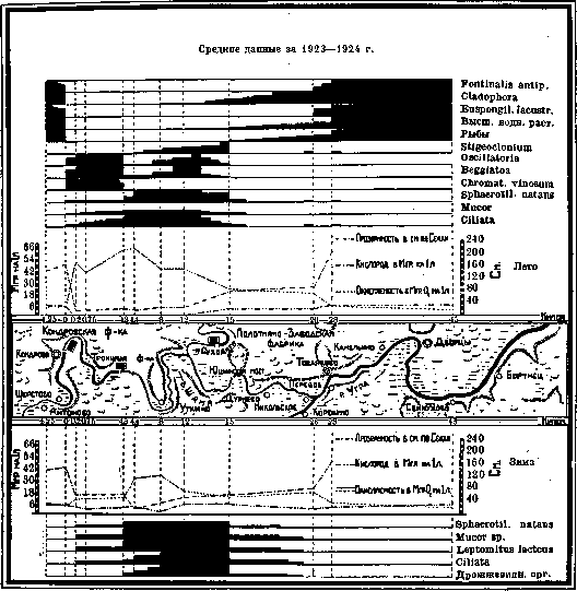
Figure 3. Diagram of the relative development of saprobic forms of benthos in the Shana and Ugra rivers under the influence of pollution by wastewater from paper mills.
Often, information about the microscopic living creatures found in the water was added to these data of microscopic analysis. The system of saprobic organisms of Kolkwitz and Marsson divides all indicator forms into 4 categories: 1) polysaprobes, 2) alpha-mesosaprobes, 3) beta-mesosaprobes, and 4) oligosaprobes. The development of polysaprobic organisms corresponds to the polysaprobic zone and indicates a very high degree of pollution with fresh, easily decomposable organic substances. In the alpha-mesosaprobic zone, characterized by alpha-mesosaprobic organisms, the pollution is either expressed more weakly or has already passed the very first stage of the self-purification process. Fresh organic compounds in it have already undergone primary decomposition. The beta-mesosaprobic zone corresponds either to even weaker pollution or to that period of the natural self-purification process in which mineralization processes are completed (complex organic substances capable of putrefaction have already been decomposed into simple mineral compounds); in it, processes of oxidation of mineral decay products take place—nitric and nitrous acids appear. The oligosaprobic zone corresponds to water that is either initially clean or in which the process of natural self-purification has been fully completed. The table (pp. 387–388) compares the main features characteristic of each zone. When performing biological analysis, methods are used to collect living material,

Figure 4. Microphotograph of plankton silk gauze No. 20 (magnified 42 times).
developed in the hydrobiology of fresh waters. Hydrobiology divides all water life into two biocenoses: plankton and benthos. Plankton includes all organisms (predominantly of microscopic size) that spend their entire life in a suspended state within the water column itself. Benthos includes all forms of plants and animals that attach themselves to the bottom (and various underwater objects) or are closely associated with it in their life. Accordingly, research methods are also divided into methods for studying plankton and methods for studying benthos. Both can be qualitative and quantitative. For collecting plankton, various kinds of nets made of silk bolting cloth are used, most often No. 20, which has mesh openings of about 70-80 μ (see Figure 4), through which the water being studied is filtered, for qualitative study in an arbitrary volume, and for quantitative study in a precisely measured volume. The organisms remaining on the walls of the net are washed into the lower part of the net, from where they are transferred with the remnants of water into a vial with a stopper and, if necessary, fixed by adding a few drops of formalin. Recently, cylindrical nets have found increasingly wide application, e.g., the Langhans zeppelin net (see Figure 5), which indeed offers great advantages. When using bolting cloth nets, only particles (living and dead) with a diameter greater than 70-80 μ are retained; Kolkwitz proposed the name seston for them. Finer particles, and among them organisms that sometimes develop very abundantly in water (nanoplankton, or dwarf plankton), pass through the net and are lost. Therefore, in addition to net plankton, it is absolutely necessary to take water samples in their natural state in order to be able to collect and study small forms as well. From the taken water samples, the organisms contained in them can be isolated by means of centrifugation (Lohmann), sedimentation (Greenberg), or filtration through special membrane filters (Kolkwitz). Figures 6 and 7 in comparison with Figure 4
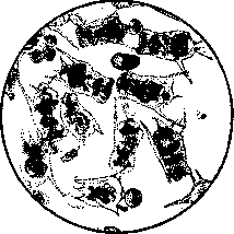
Figure 6. Microphotograph of net plankton—Brachionus pala-amphiceros. Bolting cloth No. 20 (magnified 42 times). (all images were taken at the same magnification) show clearly how substantial the difference is between net plankton and sedimentary plankton. They show what a significant part of the plankton passes (and by its size cannot help but pass) through the net. Often, one also resorts to direct study of the "natural" sample, without any
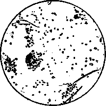
Figure 7. Microphotograph of sedimentary plankton—Euglena (magnified 42 times).
concentration of plankton, both for qualitative and quantitative analysis (in the Kolkwitz chamber, see Figure 8). For collecting benthos organisms, various kinds of dredges and trawls are used—cloth bags open at one end, attached to metal frames and supports; for hard ground, heavier ones are used, and for soft ground, lighter ones (see Figure 9). For the quantitative accounting of the bottom population, special apparatuses—bottom samplers (Figure 10)—have recently begun to be used intensively, which cut out a specific area of ground (usually 1/10 sq. m) from the bottom with all its population. The obtained ground samples are then passed through a set of metal sieves of different diameters, on which

Figure 8. Kolkwitz chamber (somewhat reduced).

Figure 9. Ekman triangular dredge (1/10 natural size).
the larger organisms remain (by means of such an apparatus, the results shown in Figures 1 and 2 were obtained). Of great importance in Biological Analysis are various kinds of growths, coatings, films, etc., formed most often by abundant clusters of microscopically small plants (algae) and animals on the surface of various underwater objects—stones, snags, piles, buoys, piers, underwater plants, etc. They are collected with knives, tweezers, nets, and scrapers. Biological analysis in the study of water bodies is used in conjunction with chemical and bacteriological studies. Its results enliven the obtained figures of chemical analysis, illuminate them, and provide a much greater opportunity to understand the significance of their changes. Biological analysis differs from chemical and bacteriological analyses in that the latter two deal only with a specific, very small volume of water, which at the moment of sampling was in the water body at a given location. Strictly speaking, all the conclusions of the chemist and bacteriologist concern not the water of the water body in general, but only a small sample taken from it. And since the chemical composition of water in a water body and the content of bacteria in it can often undergo very significant fluctuations, it is obviously necessary, to substantiate the conclusions made on the basis of these methods, to have analyses of a sufficiently large number of samples taken at different times. The situation is different in Biological Analysis, at least in relation to indicator forms of the benthic type. Various kinds of saprobic organisms, forming coatings, films, small bushes, and other types of growths on underwater objects, respond with their development to a certain average pollution of the water washing over them. They represent living apparatuses that automatically register the average degree of pollution of the water carried by a river. The water in the river at the moment of investigation may be completely clean; but if periodic discharges of pollutants into the river occurred before this, it will inevitably be reflected in the flora and fauna of the river and will not escape the biologist's control. Another difference of Biological Analysis is that with sufficient experience of the researcher, Biological Analysis can be performed incomparably faster than chemical and bacteriological studies, and therefore it is easier to cover water bodies over large areas with its help. Therefore, Biological Analysis is especially widely used as a preliminary method, by means of which one orients oneself in the distribution of pollution in the studied water body; after such a biological reconnaissance, the development of a rational plan for further research is significantly facilitated, and the number of chemical and bacteriological analyses necessary for studying the water body is usually reduced many times over.
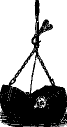
Figure 10. Petersen bottom sampler
...lowered onto an area of 1/10 sq. m of the bottom surface, in the open position (1/10 of natural size). Saprobic system (the most important indicator forms of aquatic plants and animals used by biological analysis). Acineta, an infusorian from the order of suctorians (Suctoria); a predator feeding on other infusorians, which it catches with special contracting suckers and then sucks out. It belongs to the beta-mesosaprobes. Actinophrys sol Ehrb. (see Table II, Fig. 17), an animal organism from the order of heliozoans (Heliozoa). Pseudopodia in the form of rays. It is found, predominantly, in polluted waters and belongs to the mesosaprobes. Alona, a genus of crustaceans from the order Cladocera (water fleas). It is found in clean waters and belongs to the oligosaprobes. Amphileptus Claparedli Stein, a ciliated infusorian (Ciliata) from the order of holotrichs (Holotricha), feeding, predominantly, on bacteria. Its development in water indicates an abundant development of bacteria in the latter. It belongs to the alpha-mesosaprobes. Anabaena (see Table IV, Fig. 1), an alga from the order of blue-green algae (Cyanophyceae) in the form of a curved filament composed of rounded cells, with heterocysts between them. It is common in the plankton of our standing and flowing water bodies. Sometimes it causes water blooming, which is why one of the species of this genus received the name A. flos-aquae. It is found only in clean water and belongs to the oligosaprobes. It gives the water a grassy smell. Ancylus, a small freshwater mollusk (from the gastropods—Gastropoda), having a shell in the form of a pointed (conical), cap-like shape bent at the apex. It belongs to the oligosaprobes. Anodonta, the swan mussel, a river shell—a large freshwater mollusk with a bivalve layered shell, lined from the inside with a nacreous layer. Along with the painter's mussel (Unio), it is very common in fresh waters. It lives by crawling along the bottom, burying the anterior end of the shell and the muscular foot emerging from it into silt, sand, or clay. It feeds on plankton, catching it from the water driven through the body by the inlet and outlet "siphons" located at the posterior end of the body. It belongs to the oligosaprobes. Anthophysa vegetans Butsch. (see Table II, Fig. 18), a freshwater infusorian from the order of flagellates (Flagellata). A. veg. is a colonial flagellate; its individuals, equipped with flagella, sit on the tips of a branched stalk. It is remarkable in that abundant deposits of iron oxide compounds are usually observed in its stalks, coloring them brown, similar to the sheaths of iron bacteria filaments. Its development in water indicates the presence of iron salts. Anuraea (see Table III, Figs. 15 and 16), a genus of rotifers (Rotatoria). Planktonic organisms A. aculeata Ehrb. and A. cochlearis Gosse are beta-mesosaprobes. Aphanizomenon flos-aquae Ralfs (see Table III, Fig. 1), an alga from the order of blue-green algae (Cyanophyceae), in the form of multicellular straight filaments with heterocysts and large oval "spores"; the filaments usually stick together in groups, forming flakes, which often cause the blooming of lakes, ponds, and other standing and slowly flowing waters, giving the water a greenish color. In the saprobic system, it stands among the beta-mesosaprobes. Apodya lactea (Ag) Cornu = Leptomitus lacteus Ag. (see Table II, Fig. 5), an aquatic fungus from the order Phycomycetes, or algal fungi. Its unicellular, non-septate mycelium bears extremely characteristic for Apodya constrictions (narrowings) of the filament at certain intervals, by which this fungus is easily distinguished under a microscope from other aquatic fungi. Less characteristic is the presence in its hyphae, usually near the constrictions, of large spherical inclusions, so-called cellulose bodies (Zellulinkorper). Apodya develops only in waters heavily polluted with organic substances, in the form of grayish-white or dirty mucous flakes, the size of a fist or larger, attaching to all kinds of underwater objects and sometimes covering the entire bottom and banks with a solid carpet. A very important indicator form for assessing water pollution in biological analysis. It belongs to the alpha-mesosaprobes. Arcella vulgaris Ehrb. (see Table III, Fig. 26), a rhizopod (Rhizopoda) with a protoplast enclosed in a round, cup-shaped, chitinous shell, brown in color, with an opening on the underside through which pseudopodia are extended; it is a beta-mesosaprobe. Arthrospira Jenneri-Stitz (see Table II, Fig. 8), an alga from the order of blue-green algae (Cyanophyceae), its body has the appearance of a spirally curved, with a large number of turns, motile multicellular filament of bright blue-green color. It is often found in polluted waters together with the sulfur bacterium Beggiatoa and Oscillatoria and belongs to the polysaprobes and alpha-mesosaprobes. Asellus aquaticus (L.) O.F.M., the water louse. A very common, fairly large (up to 1.5 cm) crustacean in fresh waters. It belongs to the alpha-mesosaprobes. Aspidisca (see Table III, Fig. 22), a ciliated infusorian from the order of hypotrichs, developing in polluted waters; it belongs to the beta-mesosaprobes and partly to the alpha-mesosaprobes. Asplanchna (see Table III, Fig. 12), a planktonic rotifer of fresh waters, distinguished by the transparency of its sac-like body. It belongs to the beta-mesosaprobes. Asterionella (see Table IV, Fig. 6 e), a planktonic diatom alga, the rod-shaped elements of which are arranged like the spokes of an umbrella, and a very thin film of mucus is stretched between them; something like a parachute is obtained, which allows the alga to float in the water without sinking to the bottom. It often takes part in water blooming. It is found in clean waters and belongs to the oligosaprobes. It gives the water an aromatic, sometimes fishy smell. Batrachospermum, an alga from the red algae (Florideae), in the form of a whorled, very mucous bush a few cm in height. It is common in peaty rivers. Oligosaprobe. Beggiatoa (see Table I, Fig. 12), one of many genera of sulfur bacteria, forms long, motile filaments consisting of individual cells containing inclusions of mineral sulfur. Most often, by the intertwining of its filaments, it forms whitish, very fragile films on the surface of silt. It develops only in waters containing simultaneously H2S and oxygen. Microaerophile. Its development with certainty indicates the presence of H2S in the water. And since, with the exception of sulfur springs, waters heavily polluted are usually distinguished by the content of H2S, practically Beggiatoa is an indicator of heavy water pollution and belongs to the polysaprobes. Bodonidae, flagellate forms from the genus Bodo, with two flagella, very common in heavily polluted waters. There are many species, most of which belong to the polysaprobes and mesosaprobes. Brachionus, one of the genera of rotifers (Rotatoria-Rotifera) common in fresh waters; some species develop in the plankton of polluted waters and belong to the alpha-mesosaprobes. Carchesium Lachmanni Kent. (see Table IV, Fig. 16), a sessile, colonial infusorian from the peritrichs. Individuals sit on the tips of an abundantly branched stalk, the topmost branches of which are contractile and can curl into a spiral. A bacteria-eater. In the alpha-mesosaprobic zone, it sometimes forms solid, even, white coatings on all underwater objects. An important indicator form. Chara, a genus of algae from the family Characeae. The most highly developed organisms among algae. They have the appearance of graceful little fir trees. Very common in lakes, not rare in peat waters. Oligosaprobes. Chironomus—Ch. plumosus L., the bloodworm, bright red larvae of a special type of non-biting midge living in silt. It is found in masses in polluted water bodies. Alpha-mesosaprobe. A favorite food for fish raised in aquariums, and good bait for fishing with a rod. Cladophora (see Table IV, Fig. 2), a genus of green filamentous algae (Confervales), in the form of more or less branched, usually attached bushes from a few to many cm long. Cl. crispata Ktz.—beta-mesosaprobe; other species—oligosaprobes. Closterium (see Table III, Fig. 3), a genus of algae from the family Desmidiaceae. The body shape is in the form of a crescent moon. The chromatophore is green. Sexual reproduction by conjugation. Cl. acerosum Ehrb. and Cl. moniliferum Ehrb. belong to the beta-mesosaprobes, Cl. lunula, Cl. Ehrenbergii Meneg., and others—to the oligosaprobes. Colpidium, a ciliated infusorian from the holotrichs. Very common in heavily polluted waters. A typical alpha-mesosaprobe. A bacteria-eater. Conferva, a genus of filamentous green algae (Confervales). C. bombycina—beta-mesosaprobe. Corethra (Sayomyia), an aquatic larva of a special type of midge from the chaoborids. One of the species is common for the deep layers of water of our lakes. The larva is distinguished by its transparency and the presence of swimming bladders in the body. Oligosaprobe. Dreissena (see Table IV, Fig. 12), a sessile (with a firmly attached shell) mollusk from the bivalves (Lamellibranchiata) with a bivalve shell. A Caspian immigrant, gradually spreading up rivers. It is found in large quantities in the rivers of the southern zone of the USSR, but also reaches the central regions. Oligosaprobe. Elodea canadensis R. et M., a flowering, monocotyledonous, aquatic plant, introduced to our continent from America and widely spread among us. Beta-mesosaprobe.
Eristalis tenax L., the rat-tailed maggot, a large, soft, white-gray fly larva found in very dirty water (often in masses in cesspools). It possesses a long respiratory tail of telescopic construction. Polysaprobic. Euglena (see Table I, Fig. 15), a genus of chlorophyll-bearing flagellates (Flagellata). Of the many species of the genus, E. viridis Ehrb. is especially important. A single-celled organism with one flagellum at the anterior end of the body and a red eyespot. It develops during heavy pollution, often in enormous quantities, giving the water a bright green color (blooming). Polysaprobic. Fontinalis, one of the aquatic mosses (Bryophyta), forming large dark green bushes, especially in fast currents in clean streams. Oligosaprobic. Glaucoma scintillans Ehrb., a ciliated infusorian (Ciliata) from the holotrichous ciliates (Holotricha). A bacteria-eater. Alpha-mesosaprobic. Hydra fusca L., a freshwater polyp from Hydroidea. The multicellular body forms a long, soft, contractile sac that attaches at the lower end, with a mouth opening at the upper end. Around the mouth opening sit very extensible and long tentacles, equipped with stinging organs. The tentacles catch various small animals. Oligosaprobic, but is also encountered in the beta-mesosaprobic zone. Other species are oligosaprobic. Lamprocystis roseo-persicina Schret., a bacterium from the family Coccaceae, forming pink zoogloeal coatings on various underwater objects in heavily polluted waters. Polysaprobic. Melosira varians Ag. (see Table III, Fig. 9), a filamentous alga from the diatoms (Bacillariales). A planktonic form. Belongs to the beta-mesosaprobes. Mucor, a simple aquatic fungus from the Zygomycetes. A very important indicator form, developing in heavily polluted waters, often in enormous quantities, covering all underwater objects entirely with its coatings and flakes. Characterized by the absence of septa in its hyphae; the mycelium is single-celled. Reproductive organs are not observed during development in water. Alpha-mesosaprobic. Oscillatoria (see Table II, Figs. 9 and 10), a genus of benthic blue-green algae (Cyanophyceae) from the family Oscillatoriaceae. Blue-green multicellular filaments possessing a crawling and oscillating movement. In water bodies, they form blue-green coatings on the bottom and underwater objects, often lifted to the water surface during the day by gases in the form of blue-green muddy cakes floating on the water. Some species are alpha-mesosaprobic (O. princeps Vauch., O. tenuis Ag., etc.), others are beta-mesosaprobic (O. limosa Ag.), and others are oligosaprobic (O. Agardhii Gom.). Paramaecium (see Table II, Fig. 13), the slipper animalcule, from the holotrichous infusorians. The body resembles a slipper in shape, is covered on all sides with cilia, and has a mouth opening, an anal pore, and two complex pulsating vacuoles. They feed mainly on bacteria. In heavily polluted waters, they are often found in enormous quantities. Important indicator forms, especially the polysaprobic P. putrinum Cl. et L. and the mesosaprobic P. caudatum Ehrb. Pediastrum (see Table III, Fig. 4), a genus of protococcoid algae; colonies in the form of a rounded plate, passively floating in the water. One of the most common planktonic algae of fresh waters. P. Boryanum Menegh. is alpha-mesosaprobic. Other species are oligosaprobic. Plumatella, a genus of bryozoans (Bryozoa), colonial animals that sometimes form very large colonies, the size of a fist (Pl. fungosa Pall.). In clean and slightly polluted waters. Beta-mesosaprobic and oligosaprobic. Polytoma uvella, a colorless alga from the Protococcales, with two flagella at the anterior end of the body. In heavily polluted waters, often in enormous quantities. An important indicator form. Polysaprobic. Psychoda, small flies (Diptera), the larvae of which live in heavily polluted water; they often develop in enormous quantities in the body of biological oxidizers. Rotifer (see Table II, Fig. 14), a genus of rotifers (Rotatoria), multicellular animals of the benthic type. Especially important are R. vulgaris Schr. and R. neptunius Ehrb. (actinurus), which live in dirty waters; both are alpha-mesosaprobic. Sphaerotilus (see Table II, Fig. 6), a very important indicator form of biological analysis, belongs to the filamentous bacteria (Trichobacteriaceae). In heavily polluted waters, it forms large flakes, reaching many centimeters, consisting of filaments; each filament is formed from many individual cylindrical cells and is covered by a common sheath. The common form is Sph. natans Ktz. and the rarer, pink, Sph. roseus Zopf. Both are polysaprobic. Spirogyra, a genus of algae from the Conjugatae. Forms long (centimeters and even meters) filaments, more often free-floating, less often attached. Chromatophores are in the form of green ribbons, curved spirally along the cells. Sexual reproduction by conjugation. Very common in our waters. Sp. crassa Ktz. and Sp. porticalis Cleve are beta-mesosaprobic; other species are oligosaprobic. Stentor (see Table III, Fig. 23), the trumpet animalcule, a genus of ciliated infusorians (Ciliata), one of the largest infusorians, often easily distinguishable by the naked eye. St. coeruleus Ehrb. and St. roeseli Ehrb. are beta-mesosaprobic; St. polymorphus Ehrb. and others are alpha-mesosaprobic. Stigeoclonium (see Table II, Fig. 3), a genus of green algae (Chlorophyceae) from the family Chaetophoraceae. Forms green, delicate, highly branched bushes. St. tenue Kg. is alpha-mesosaprobic. Tubifex (see Table I, Fig. 17), a genus of bristle worms (Oligochaeta). In body structure, it is close to the earthworm. Lives in silt. Size 1-4 cm. T. tubifex O. F. M. is in dirty waters, alpha-mesosaprobic, and is also encountered in the polysaprobic zone. Volvox (see Table IV, Fig. 13), a genus of colonial algae from the family Volvocaceae, in the form of a hollow ball up to the size of a pinhead, with a mass of flagella extending in pairs from each individual. The ball leads a planktonic lifestyle, swimming in the water with the help of flagella. All species of this genus are oligosaprobic. It gives the water a fishy odor. Vorticella (see Table I, Fig. 18), the bell animalcule, a genus of sessile ciliated infusorians (Ciliata), belonging to the peritrichous ciliates. The single-celled body is in the form of a bell, sitting on a contractile long stalk. On the upper flat end of the body is the peristome with cilia arranged in a spiral. By the movement of the cilia, the peristome drives small particles floating in the water into the mouth opening located in the center. V. microstoma Ehrb. and V. putrina O. F. M. are polysaprobic; V. convallaria Ehrb. is alpha-mesosaprobic; V. campanula Ehrb. and others are beta-mesosaprobic; V. nebulifera Ehrb. is oligosaprobic. Zoogloea ramigera Itzig. (see Table I, Fig. 1), a bacterium from the family Bacteriaceae, forming branched zoogloeae of a peculiar shape in heavily polluted waters. Polysaprobic.
Related articles
Mentioned in
Cite this page
“Biological Analysis.” Soviet Medical Encyclopedia. English translation of Bolshaya Meditsinskaya Entsiklopediya, 1st ed. (Moscow, 1928–1936), ed. N. A. Semashko. https://sovietmedicalencyclopedia.pages.dev/article/biological-analysis/