Poisonous Animals
Historical document, translated for reference. It reflects medical knowledge of the 1920s–30s and is not medical advice.
Summary
This article from the 1928-1936 Soviet Medical Encyclopedia distinguishes between truly poisonous and accidentally poisonous animals, examines their toxic properties, organs, and effects on humans and other animals.
Encyclopedia article (1928–1936)
Poisonous animals are characterized by the constant or periodic presence in their organisms of substances possessing poisonous properties in relation to beings of other species. It is necessary to distinguish truly poisonous animals from accidentally poisonous creatures, which individual specimens of harmless species may become due to various circumstances. Examples of true poisonous animals include the bee, scorpions, the adder, and others. Individual specimens of red fish (sturgeon, etc.) may also acquire toxic properties; such "accidentally" poisonous fish can cause fatal poisoning in humans through the action of "fish poison." Comparative toxicological experiments provide abundant material for judging the relativity of poisonous properties, since different animal species may react differently to the same poisonous substance. Strychnine is a deadly poison for mammals, while the rhinoceros bird feeds on the seeds of Nux vomica; the sting of bees is very painful for humans, horses, and other animals, while the frog and the bee-eater bird excellently devour bees. The first moment determining poisoning by poisonous animals is the dose of poison introduced into the organism. Not every adder bite ensures the introduction of the same amount of poison. Here plays a role not only the age of the snake (a small adder of course gives less poison than a large one), but even such a fact as the duration of the preceding period of rest of the poison glands. Various states of poisonous animals can influence the production of poison. For example, the cobra becomes ten times more poisonous after molting than before it. The place where the poison enters the organism has an important influence on the outcome of poisoning, i.e., that barrier which the poison encounters. The intact human skin is impermeable to snake venom. When its integrity is violated (snake bite, prick of the tip of a poisoned arrow), the poison enters the blood and gives its characteristic pharmacological effect. The same poison, when swallowed and entering the stomach, has no effect, as it is neutralized when passing through the liver barrier. But even with the typical entry of poison into the organism, the results may be different. If the poison enters a place poor in blood vessels, its absorption is slower, which also affects the general reaction of the organism up to the manifestation of resistance to poisoning, completely unrelated to immune properties. In particular, pigs are resistant to the bite of the rattlesnake, which is explained by the presence of a thick layer of fat under the skin, which is poor in vessels; at the same time, there are no antitoxic substances in the blood of pigs; meanwhile, in the blood serum of the ichneumon and hedgehog, there are substances that bind snake venom to some extent. Among the features of the organism being poisoned by poisonous animals, its size (resp. age) comes to the fore (of course, besides its specific properties); therefore, as in relation to other poisons, the poisons of poisonous animals are characterized by the size of the toxic or lethal dose, calculated per 1 kg of weight of a human or some animal. Individual peculiarities of the organism in turn can influence the course and outcome of poisoning by poisonous animals. There are cases of idiosyncrasy or conversely of innate immunity or acquired insusceptibility to the poison, in particular after a successfully overcome poisoning. Such accidental circumstances as the state of intoxication and other moments cannot but influence the action of poisonous animals. Knowing how poisonous animals poison humans is practically important for finding measures to prevent possible poisonings. The poisonous properties of many poisonous animals can or may be easily discovered in natural relationships. The hornet stings a human; a human eats poisonous fish, etc. In both cases, poisoning is the result of ordinary "life" relationships, and the poisonousness of such animals can be considered obvious; at the same time, there are many facts confirming the presence of toxic properties which, under natural conditions, do not find their obvious application on the part of poisonous animals in relation to the outside world and are discovered only in experimental research. For example, the blood serum of the eel or skate (Raja) is poisonous when injected into various animals. Poisonous animals possessing this kind of poisonousness are called covertly poisonous. Poisonous animals either have special poisonous organs or various parts of their body (for example, the ovary of fish, blood, etc.) turn out to be poisonous. Poisonous organs in turn are either a modification of organs usually existing in this group of animals (for example, the salivary glands of insects, differing in the poisonousness of their sting), or they arose in the phylogenesis of poisonous animals as a "new acquisition" (for example, the poison glands of scorpions). The poisonous organs of animals consist of a poison-producing gland (poison gland) and a piercing (wounding) apparatus, by means of which the integrity of the coverings of a human or animal is violated and the poison is introduced into the thickness of the tissues (resp. into the blood); however, many poisonous animals have only poison glands, being devoid of any wounding "adaptations" (for example, ants with poison glands and a reduced sting). In this connection, they speak of "armed" and "unarmed" poisonous animals. I. Poisonous animals with poisonous organs. Even in some protozoa there are poisonous organoids. Thus, in the cortical layer of the protoplasm of various infusoria are laid down rod-shaped formations called trichocysts. With various irritations, the infusoria reflexively "shoots" out trichocysts, which pierce attacking predatory infusoria Didinium (fig. 1) and by their toxic influence can force them to drop their prey. For coelenterates, stinging, or nettling cells (fig. 2) are very characteristic, located in the ectoderm of these animals. In such a cell there is a capsule with a spiral thread turned inward. The latter when the nettling cell works is thrown outward (fig. 3) and pierces the coverings of the organism that touched the coelenterate. The latter experiences either a sensation of burning or
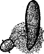
Figure 1. Paramecium, releasing a cloud of trichocysts when attacked by the predatory infusoria Didinium.
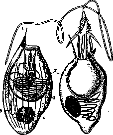
Figure 2. Nettling cells of hydra, on the right in a relaxed state: 1-kinocilium; 2-capsule; 3-supporting rods; 4-nucleus; 5-spiral thread in the protoplasm.
is paralyzed and becomes prey for the coelenterate. Thus the hydra catches small crustaceans, and actiniae capture small fish. The effect of the "burning" action is especially great when the human body comes in contact with siphonophores (Physalia); the human reaction to the poison of jellyfish can be intensified due to the state of allergy. Sponge divers, due to frequent contact with actiniae sitting on the sponges being torn off, sometimes contract a professional disease consisting in ulceration of the fingers and general intoxication. In the nettling cells
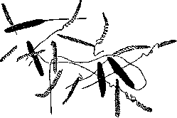
Fig.
Nettling capsules of the Black Sea Actinia equina. both the secretory part and the piercing apparatus are localized in the same cell. Animals with poison glands and a special piercing apparatus have differently constructed poisonous apparatus. To this category of poisonous animals belong: 1) stinging caterpillars of butterflies with twisted chitinous hairs, for example the fragile serrated hairs of the caterpillar of the goldtail. In the cavities of the hairs there is a poisonous secretion (fig. 4), secreted by special glandular cells of the hypodermis at the base of the hairs.
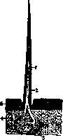
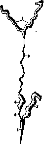
Figure 4.
FIG. 5.
Figure 4. Diagram of the structure of a poisonous hair (1) of an insect: 2-cavity of the hair with secretion; 3-glandular cell; 4-hypodermis; 5-chitin. Figure 5. Poisonous apparatus of the carpenter bee: 1-acid glands; 2-their common canal; 3-alkaline gland; 4-sting; 5-excretory duct of the reservoir; 6-reservoir. The latter are extremely fragile, easily break off even when the caterpillar moves and are carried by the wind. When inhaled, they pierce the mucous membranes of the nose, mouth, get into the eyes and cause various disorders (stomatitis, aphthae, conjunctivitis). When caterpillars are grasped with hands, they pierce the skin and cause the formation of blisters. In dry hairs, the poison can be preserved for over six
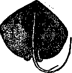
Figure 6.
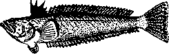
The stingray Trygon pastinaca. of seven years (E. Pavlovsky and A. Stein). Such poisonous caterpillars are also possessed by the yellow-tailed moth (Rhog-thesia similis) and the processionary moth (Thaumetopoea processionea) has gained particular fame. 2) Multicellular sac glands with a special stinger are possessed by scorpions. Their thin "postabdomen" ("tail") ends in a chitinous vesicle (ampulla), inside of which lie two poisonous glands, opening into an acute, hard, curved stinger (see Scorpions, fig. 2, B). 3) In Hymenoptera, only females possess poisonous organs (fig. 5), because the poisonous apparatus is modified parts of the female reproductive organs. Its secretory part consists of two glands—a tubular one (with a reservoir) that secretes an "acidic" secretion, and a sac-like one that produces an alkaline secretion. The degree of development of these parts varies greatly, depending on the species. The poison is a mixture of both secretions, which mix when they exit externally. These glands are homologous to the accessory glands of the female reproductive organs. The piercing part is represented by a stinger-complex of chitinous parts, which are homologs of the ovipositor. Well-known examples of poisonous Hymenoptera are the honey bee, bumblebees, wasps, etc. The lethal dose of bee venom for an adult is considered to be about 500 simultaneous stings by bees. The characteristics of bee venom are: burning local pain, swelling at the sting site, disturbances in cardiac activity, nausea, vomiting, etc. When stung by a hornet, enormous edemas develop. Bee venom has been tried for the treatment of rheumatism, lumbago, sciatica, gout, dropsy, etc., by subjecting patients to stings from one to 50-100 bees per day (with a sequential increase). Such "Bienenstichkuren" give certain results, but it is necessary to consider the side effects of treatment caused by the action of this venom. 4) In some vertebrates, there are cutaneous poisonous glands armed with a piercing apparatus in the form of bony spines or fin rays. Thus, the stingray (fig. 6) (Trygon pastinaca, Black Sea) has on its tail a powerful, laterally notched spine with two longitudinal grooves, which are filled with highly developed clusters of single-cell cutaneous glands of the deep layers of the epidermis; their secret enters the wound inflicted by the tail spine. A more perfect poisonous apparatus is possessed by some spiny-finned fishes, such as Scorpaena porcus and Trachinus draco (Black Sea) (fig. 7), Sebastes norwegicus (Murmansk), Pterois volitans, Synanceia and others. From the catfish family, a similar apparatus is possessed by Plotosus and others. The scheme of the structure of the poisonous apparatus of fish is as follows. The sharp, spiny fin rays have a pair of longitudinal grooves; in them lie spindle-shaped poisonous glands, formed by a mass of huge glandular cells secreting a serous secretion (fig. 8). Between them lie flat supporting cells (which are homologous to indifferent cells of the epidermis). Under normal conditions, such glands do not have an excretory duct, as they belong to the type of solid or compact multicellular glands. An excretory duct is formed in them during holocrine secretion, when part of the glandular cells disintegrates completely, and the framework of supporting cells is broken. The poison enters human tissue when pricked by such a fish. In the weever fish, in addition to the fin glands, there is also a pair of powerful poisonous glands on the operculum, connected with a strong spine of the gill cover.
Figure 7. Trachinus draco.
associated with a strong spine of the gill cover. o) In many animals, poisonous glands are connected with oral organs, which form their piercing part. Such are the jaws or hypopharynx of arthropods and the teeth of snakes. There are particularly many such poisonous animals among arthropods (spiders, centipedes, insects). In spiders (fig. 9), there are two tubular poisonous glands located in the cephalothorax or in the main segment of the upper jaws (chelicerae). The latter end in a movable sharp claw, at the apex of which opens a thin excretory duct of the poisonous gland. The venom is ejected from the glands under the pressure of their powerful muscular sheath. Spider venom serves to kill prey, which the spider
then sucks out. There are spiders that are very poisonous to humans, such as various species of black widows (see) and tropical spiders of Brazil, for example Lycosa raptoria, Phormictopus and others, causing a very severe local reaction (gangrenous tissue necrosis) and sometimes a fatal outcome. Tarantulas (Trochosa singoriensis, Lycosa tarentula) do not possess the degree of venom and danger attributed to them by popular opinion. The largest spiders Mygale (bird-eating spiders) are apparently not so terrifying. The bite of the spider Chiracanthium nutrix is noted to be painful. The venom of the cross spider (Epeira) has been studied in more detail. Various toxic principles have been found in extracts from spider bodies. Epeiratoxin is contained in spider eggs. When injected into dogs and cats, they begin to have convulsions, suffocation, and death during a new convulsive attack. Another principle—epeiralisin—is a hemolytic toxin; it is also contained in spider eggs and is apparently a complex system of various substances. A similar principle in other spiders is called arachnolysin. It is important to keep in mind that epeiratoxin and epeiralisin have nothing to do with the venom of the poisonous glands of spiders.
Many ticks also cause poisonous bites, for example the straw itch mite Pediculoides ventricosus (fig. 10), which sometimes contaminates grain, straw, etc. When unloading grain or when a person sleeps on straw, these mites attack him. The skin turns red and is covered with numerous nodules, often of urticarial character, with a feeling of intense itching and burning. The larvae of harvest mites (Trombicula autumnalis) cause the appearance of itchy papules ("strawberry disease") on human skin. Various flour mites (Tyroglyphidae) when working with flour, dried fruits, vanilla cause the appearance of a pruriginous rash with the formation of small, intensely itching nodules. The onion mite (Rhizoglyphus hyacinthi) also has a toxic effect on the skin. It is very likely that the cause of intoxication is the saliva of Pediculoides and Trombicula. Regarding other mites, the possibility of the action of the juices of their crushed bodies when rubbed into human skin is not excluded. The bite of the chicken mite Dermanyssus gallinae (family Gamasidae) has a significant subjective effect on humans. Many mites of the superfamily Ixodoidea occasionally attack humans. The bite of some is very painful, while others do not cause any subjective sensations despite a well-developed local reaction (Ornithodorus papillipes, Ornithodorus lahorensis, etc.). Phalanges (bichorh, solpuga) are also classified as poisonous animals, but without sufficient grounds. Their bite with powerful chelicerae, which crush the body coverings, is very painful, but phalanges have no poisonous organs or active principles (E. Pavlovsky and A. Stein). If complications from bites by phalanges do occur, it is due to the introduction of secondary infection into the wound. Part of centipedes, specifically the Chilopoda, has cylindrical poisonous glands opening at the apex of the movable claw of the gnathopod (fig. 11). Such are the centipede, the house centipede (Scutigera), which is beneficial by its energetic destruction of house flies, etc. The venomousness of centipedes has no essential significance for humans. Among insects, many species cause a bite (more precisely, a sting). Such are lice (Pediculus), the bed bug, mosquitoes, fleas, Phlebotomus, Simulium, Culicoides, horseflies, gadflies, etc. The sensitivity of different people varies. The sting of some bugs (Reduvius) is very painful. The primary reaction of human skin to the bite of different groups of insects varies. With repeated exposures,
Figure 11. Gnathopod (3) of centipede with poisonous glands (2); 1-excretory canal.
allergic phenomena are added. The effect of the poisonous sting of insects must be sharply distinguished from the painful consequences after being bitten by insects that are vectors of various disease pathogens. - Among vertebrate animals, the bite of the moray eel (Mediterranean Sea) has doubtful venomousness. The large lizard Gila monster (Mexico, California) is undoubtedly poisonous, possessing poisonous teeth in the lower jaws and submaxillary poisonous glands. Classical poisonous animals are snakes (see). Along with such undoubtedly venomous species as vipers and front-fanged snakes, "po-
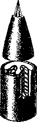
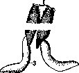
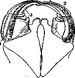
Rear-fanged snakes (Opisthoglypha) are considered 'potentially venomous,' as their venomous grooved teeth lie deep in the mouth at the back of the jaws and therefore do not produce wounds when the mouth is opened in the usual manner. Examples of rear-fanged snakes include the arrow snake (Tarbometopon lineolatum), the Caucasian cat snake (Tarbophis iberus), and others. The active principles of snake venom are echidnase or echidnin, echidnotoxin, and echidnovaccine (Phisalix). They differ in their resistance to heat. Echidnotoxin withstands brief boiling. It is a paralytic venom; whereas echidnase causes local action (hemorrhagic edema, cytolysis). Echidnovaccine has vaccinating properties with respect to snake venom. Animals with venomous glands but without a piercing apparatus exist among both vertebrates and invertebrates. The venomous secret of such animals can act on the skin coverings, mucous membranes, respiratory and olfactory organs. 1) The skin glands of amphibians have been most studied in relation to their venom. The glands themselves are simple sacs immersed in the corium (Fig. 12); from the basal membrane lie ectodermal cells resembling smooth muscle elements; on them rest large glandular cells. Granular, serous, and mucous skin glands are distinguished. The first are particularly large: powerful clusters of them in the form of parotids lie behind the eyes of the toad, salamander, and newt. From them a milky-like secret can be ejected. In addition, in the Surinam pipa the glands are arranged in longitudinal lines along the length of the body on the back and closer to the sides. The difference between the two types of glands is relative, as in the same gland there can be both mucous and granular cells simultaneously. Venomous glands quite reliably protect some amphibians from enemies. Thus, toads have almost no natural enemies (of course, excluding humans). The venom of toads is bitter and nauseating in taste. From it, various researchers have isolated active principles—phrynin, bufidin, and bufotoxin. A detailed pharmacological study of toad venom was carried out by N. P. Kravkov. The main toxic properties are inherent in the secret of the granular glands. Interestingly, toad venom can replace digitalis and cocaine in the nature of its action. Staderini even performed eye operations under toad venom instead of cocaine. This coincides with the use of toads for treating toothache in folk medicine (toads are tied to the gums). However, such treatment sometimes ends in fatal poisoning (Argentina), since mucous membranes, unlike the skin coverings, absorb the venom of amphibians. The salamander (Salamandra maculosa) was also an object of venom study, in which samarin and samaridin were discovered (Faust). Pharmacologically, the venom of the salamander belongs to the group of convulsive poisons. The venomousness of the salamander has no practical significance for humans, but the venom of some tailless amphibians, such as the spotted poison frog (Dendrobates tinctorius), in South America is used by savages for poisoning arrow tips. 2) Among arthropods, various insects that se strongly smelling, rapidly evaporating liquids have their own kind of unarmed venomous glands. The actual toxic properties of these products have not been studied, and the classification of corresponding animals as venomous is conditional. Such are darkling beetles, for example, blaps with fragrant glands; blister beetles with anal glands, stink bugs with fragrant glands, and many others. Insects possessing the phenomenon of blood-squirting, when toxic blood is secreted or ejected outward through special openings in the body (for example, in the joints of the limbs), should also be included in the category of unarmed venomous apparatus; such property is possessed by blister beetles, ladybugs, and others. Unarmed fragrant glands are also found in some centipedes (for example, Fontaria gracilis). Unarmed venomous glands are also called protective glands, and often a bright body coloration of their owner is associated with them, which is interpreted as 'warning' or 'cautioning.' However, in many cases, owners of such coloration still become prey to other animals, therefore the indicated interpretation is not firmly substantiated (Heikertinger). II. Animals without special venomous apparatus, possessing toxic properties due to the venomousness of some of their organs, tissues, or excretory products. 1) Some animals are venomous when consumed as food or generally when taken per os. Such are, for example, poisonous fish causing poisoning called ciguatera (not to be confused with poisoning from fish venom). In this regard, various fused-jaw fishes (Plectognathi), pufferfish, tetradons, and four-toothed fish are most well-known: Tetrodon (Spheroides) pardalis (Fig. 13), Tetrodon porphyreus, Tetrodon inermis, and many others. They live in tropical seas, there are many of them in Japan. Near Vladivostok, Tetrodon vermicularis and others are found. The meat of these fish is edible, but their ovaries and roe are very poisonous. In 22 years in Japan, 3106 cases of pufferfish poisoning were recorded with 2090 fatal outcomes. The venom of tetradons (or fugu) acts on the central nervous system, paralyzing the respiratory or vasomotor centers with parallel paralysis of the peripheral endings of motor nerves. Tahara isolated tetrodonin and tetrodotoxin (= tetrodonic acid) from the roe of fresh pufferfish. The toxic principle is tetrodotoxin. Ostracion, or trunkfish, is also considered poisonous in Japan. Among other bony fishes, various species of the Central Asian Schizothorax (Fig. 14) are poisonous. Their meat is tasty, but their roe (ovaries) and supposedly the black peritoneal membrane have toxic properties and cause vomiting, diarrhea, severe exhaustion, and other phenomena. It should be noted that there are no detailed descriptions of cases of human poisoning by marinka in the medical literature. Kushelevsky notes that its roe does not lose its poisonous properties from salting and cooking. The barbel (Barbus) (Fig. 15), widely distributed in central and southern Europe, is poisonous mainly during the breeding season. Eating its roe causes a cholera-like disease (Wagenbrenner). Among tropical fishes, Clupea (Meletta) venenosa and Clupea (Meletta) trissa are considered very poisonous, and cases of fatal poisoning after consuming them are noted in the literature. With respect to cyclostome fishes, there are indications of cases of severe poisoning after eating soup from river lampreys (Lampetra planeri). It is believed that the venom is localized in the secret of unicellular skin glands, therefore for safety it is recommended to salt fresh lampreys and thoroughly wash off the abundant mucous secret that is released at this time. In some southern coastal areas, sea urchins, specifically their gonads, are eaten. It has long been noticed that during the sexual activity period, the ovaries of Toxopneustes lividus become poisonous. 2) When certain insects are taken per os (for example, when taken internally as an aphrodisiac, blister beetles—Lytta, Mylabris, and others), severe poisoning develops with kidney damage (action of cantharidin). 3) Tissue venomousness is possessed by such insects as beetles of the genus Paederus (family Staphylinidae); their blood and internal genitalia contain a toxic principle and cause serious bullous dermatitis on the skin, not identical to the action of Spanish flies. For the action to manifest, the beetle must be crushed on the skin and its juices must get into scratches on the epidermis. Even more serious consequences are observed when these beetles get into the eye and are crushed in it [conjunctivitis, inflammation of the eyelid thickness, inflammation of the cornea and iris, etc. (E. Andresen)]. The larvae of the beetle Diamphidia locusta (tropical Africa) contain a very strong poison in their juices, which the Bushmen use for poisoning arrow tips. The adult beetle, however, is completely harmless. 4) The toxic properties of various endoparasites must be specially considered. Living in the body of the host, they cause its chronic intoxication. The nature of their active principles varies. Some are exotoxins, i.e., they are secreted during the life of the parasite from its body as a secret of special glands (for example, in hookworms; the horse nematode Sclerostomum secretes sclerototoxin in the secret of its head glands, which has hemolytic properties and dissolves the cells of the intestinal mucous membrane) or in the form of excretory products. Other active principles are released only
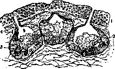
Figure 12. Section of frog skin with alveolar glands: 1-epidermis; 2-oxophilic glandular cells; 3-mucous glandular cells; 4-excretory duct.
fragrant glands are also found in some centipedes (for example, Fontaria gracilis). Unarmed venomous glands are also called protective glands, and often a bright body coloration of their owner is associated with them, which is interpreted as 'warning' or 'cautioning.' However, in many cases, owners of such coloration still become prey to other animals, therefore the indicated interpretation is not firmly substantiated (Heikertinger). II. Animals without special venomous apparatus, possessing toxic properties due to the venomousness of some of their organs, tissues, or excretory products. 1) Some animals are venomous when consumed as food or generally when taken per os. Such are, for example, poisonous fish causing poisoning called ciguatera (not to be confused with poisoning from fish venom). In this regard, various fused-jaw fishes (Plectognathi), pufferfish, tetradons, and four-toothed fish are most well-known: Tetrodon (Spheroides) pardalis (Fig. 13), Tetrodon porphyreus, Tetrodon inermis, and many others. They live in tropical seas, there are many of them in Japan. Near Vladivostok, Tetrodon vermicularis and others are found. The meat of these fish is edible, but their ovaries and roe are very poisonous. In 22 years in Japan, 3106 cases of pufferfish poisoning were recorded with 2090 fatal outcomes. The venom of tetradons (or fugu) acts on the central nervous system, paralyzing the respiratory or vasomotor centers with parallel paralysis of the peripheral endings of motor nerves. Tahara isolated tetrodonin and tetrodotoxin (= tetrodonic acid) from the roe of fresh pufferfish. The toxic principle is tetrodotoxin. Ostracion, or trunkfish, is also considered poisonous in Japan. Among other bony fishes, various species of the Central Asian Schizothorax (Fig. 14) are poisonous. Their meat is tasty, but their roe (ovaries) and supposedly the black peritoneal membrane have toxic properties and cause vomiting, diarrhea, severe exhaustion, and other phenomena. It should be noted that there are no detailed descriptions of cases of human poisoning by marinka in the medical literature. Kushelevsky notes that its roe does not lose its poisonous properties from salting and cooking. The barbel (Barbus) (Fig. 15), widely distributed in central and southern Europe, is poisonous mainly during the breeding season. Eating its roe causes a cholera-like disease (Wagenbrenner). Among tropical fishes, Clupea (Meletta) venenosa and Clupea (Meletta) trissa are considered very poisonous, and cases of fatal poisoning after consuming them are noted in the literature. With respect to cyclostome fishes, there are indications of cases of severe poisoning after eating soup from river lampreys (Lampetra planeri). It is believed that the venom is localized in the secret of unicellular skin glands, therefore for safety it is recommended to salt fresh lampreys and thoroughly wash off the abundant mucous secret that is released at this time. In some southern coastal areas, sea urchins, specifically their gonads, are eaten. It has long been noticed that during the sexual activity period, the ovaries of Toxopneustes lividus become poisonous. 2) When certain insects are taken per os (for example, when taken internally as an aphrodisiac, blister beetles—Lytta, Mylabris, and others), severe poisoning develops with kidney damage (action of cantharidin). 3) Tissue venomousness is possessed by such insects as beetles of the genus Paederus (family Staphylinidae); their blood and internal genitalia contain a toxic principle and cause serious bullous dermatitis on the skin, not identical to the action of Spanish flies. For the action to manifest, the beetle must be crushed on the skin and its juices must get into scratches on the epidermis. Even more serious consequences are observed when these beetles get into the eye and are crushed in it [conjunctivitis, inflammation of the eyelid thickness, inflammation of the cornea and iris, etc. (E. Andresen)]. The larvae of the beetle Diamphidia locusta (tropical Africa) contain a very strong poison in their juices, which the Bushmen use for poisoning arrow tips. The adult beetle, however, is completely harmless. 4) The toxic properties of various endoparasites must be specially considered. Living in the body of the host, they cause its chronic intoxication. The nature of their active principles varies. Some are exotoxins, i.e., they are secreted during the life of the parasite from its body as a secret of special glands (for example, in hookworms; the horse nematode Sclerostomum secretes sclerototoxin in the secret of its head glands, which has hemolytic properties and dissolves the cells of the intestinal mucous membrane) or in the form of excretory products. Other active principles are released only
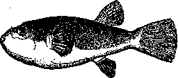
Figure 13. Spheroides chrysops.
fragrant glands are also found in some centipedes (for example, Fontaria gracilis). Unarmed venomous glands are also called protective glands, and often a bright body coloration of their owner is associated with them, which is interpreted as 'warning' or 'cautioning.' However, in many cases, owners of such coloration still become prey to other animals, therefore the indicated interpretation is not firmly substantiated (Heikertinger). II. Animals without special venomous apparatus, possessing toxic properties due to the venomousness of some of their organs, tissues, or excretory products. 1) Some animals are venomous when consumed as food or generally when taken per os. Such are, for example, poisonous fish causing poisoning called ciguatera (not to be confused with poisoning from fish venom). In this regard, various fused-jaw fishes (Plectognathi), pufferfish, tetradons, and four-toothed fish are most well-known: Tetrodon (Spheroides) pardalis (Fig. 13), Tetrodon porphyreus, Tetrodon inermis, and many others. They live in tropical seas, there are many of them in Japan. Near Vladivostok, Tetrodon vermicularis and others are found. The meat of these fish is edible, but their ovaries and roe are very poisonous. In 22 years in Japan, 3106 cases of pufferfish poisoning were recorded with 2090 fatal outcomes. The venom of tetradons (or fugu) acts on the central nervous system, paralyzing the respiratory or vasomotor centers with parallel paralysis of the peripheral endings of motor nerves. Tahara isolated tetrodonin and tetrodotoxin (= tetrodonic acid) from the roe of fresh pufferfish. The toxic principle is tetrodotoxin. Ostracion, or trunkfish, is also considered poisonous in Japan. Among other bony fishes, various species of the Central Asian Schizothorax (Fig. 14) are poisonous. Their meat is tasty, but their roe (ovaries) and supposedly the black peritoneal membrane have toxic properties and cause vomiting, diarrhea, severe exhaustion, and other phenomena. It should be noted that there are no detailed descriptions of cases of human poisoning by marinka in the medical literature. Kushelevsky notes that its roe does not lose its poisonous properties from salting and cooking. The barbel (Barbus) (Fig. 15), widely distributed in central and southern Europe, is poisonous mainly during the breeding season. Eating its roe causes a cholera-like disease (Wagenbrenner). Among tropical fishes, Clupea (Meletta) venenosa and Clupea (Meletta) trissa are considered very poisonous, and cases of fatal poisoning after consuming them are noted in the literature. With respect to cyclostome fishes, there are indications of cases of severe poisoning after eating soup from river lampreys (Lampetra planeri). It is believed that the venom is localized in the secret of unicellular skin glands, therefore for safety it is recommended to salt fresh lampreys and thoroughly wash off the abundant mucous secret that is released at this time. In some southern coastal areas, sea urchins, specifically their gonads, are eaten. It has long been noticed that during the sexual activity period, the ovaries of Toxopneustes lividus become poisonous. 2) When certain insects are taken per os (for example, when taken internally as an aphrodisiac, blister beetles—Lytta, Mylabris, and others), severe poisoning develops with kidney damage (action of cantharidin). 3) Tissue venomousness is possessed by such insects as beetles of the genus Paederus (family Staphylinidae); their blood and internal genitalia contain a toxic principle and cause serious bullous dermatitis on the skin, not identical to the action of Spanish flies. For the action to manifest, the beetle must be crushed on the skin and its juices must get into scratches on the epidermis. Even more serious consequences are observed when these beetles get into the eye and are crushed in it [conjunctivitis, inflammation of the eyelid thickness, inflammation of the cornea and iris, etc. (E. Andresen)]. The larvae of the beetle Diamphidia locusta (tropical Africa) contain a very strong poison in their juices, which the Bushmen use for poisoning arrow tips. The adult beetle, however, is completely harmless. 4) The toxic properties of various endoparasites must be specially considered. Living in the body of the host, they cause its chronic intoxication. The nature of their active principles varies. Some are exotoxins, i.e., they are secreted during the life of the parasite from its body as a secret of special glands (for example, in hookworms; the horse nematode Sclerostomum secretes sclerototoxin in the secret of its head glands, which has hemolytic properties and dissolves the cells of the intestinal mucous membrane) or in the form of excretory products. Other active principles are released only
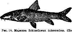
(Berg.)
fragrant glands are also found in some centipedes (for example, Fontaria gracilis). Unarmed venomous glands are also called protective glands, and often a bright body coloration of their owner is associated with them, which is interpreted as 'warning' or 'cautioning.' However, in many cases, owners of such coloration still become prey to other animals, therefore the indicated interpretation is not firmly substantiated (Heikertinger). II. Animals without special venomous apparatus, possessing toxic properties due to the venomousness of some of their organs, tissues, or excretory products. 1) Some animals are venomous when consumed as food or generally when taken per os. Such are, for example, poisonous fish causing poisoning called ciguatera (not to be confused with poisoning from fish venom). In this regard, various fused-jaw fishes (Plectognathi), pufferfish, tetradons, and four-toothed fish are most well-known: Tetrodon (Spheroides) pardalis (Fig. 13), Tetrodon porphyreus, Tetrodon inermis, and many others. They live in tropical seas, there are many of them in Japan. Near Vladivostok, Tetrodon vermicularis and others are found. The meat of these fish is edible, but their ovaries and roe are very poisonous. In 22 years in Japan, 3106 cases of pufferfish poisoning were recorded with 2090 fatal outcomes. The venom of tetradons (or fugu) acts on the central nervous system, paralyzing the respiratory or vasomotor centers with parallel paralysis of the peripheral endings of motor nerves. Tahara isolated tetrodonin and tetrodotoxin (= tetrodonic acid) from the roe of fresh pufferfish. The toxic principle is tetrodotoxin. Ostracion, or trunkfish, is also considered poisonous in Japan. Among other bony fishes, various species of the Central Asian Schizothorax (Fig. 14) are poisonous. Their meat is tasty, but their roe (ovaries) and supposedly the black peritoneal membrane have toxic properties and cause vomiting, diarrhea, severe exhaustion, and other phenomena. It should be noted that there are no detailed descriptions of cases of human poisoning by marinka in the medical literature. Kushelevsky notes that its roe does not lose its poisonous properties from salting and cooking. The barbel (Barbus) (Fig. 15), widely distributed in central and southern Europe, is poisonous mainly during the breeding season. Eating its roe causes a cholera-like disease (Wagenbrenner). Among tropical fishes, Clupea (Meletta) venenosa and Clupea (Meletta) trissa are considered very poisonous, and cases of fatal poisoning after consuming them are noted in the literature. With respect to cyclostome fishes, there are indications of cases of severe poisoning after eating soup from river lampreys (Lampetra planeri). It is believed that the venom is localized in the secret of unicellular skin glands, therefore for safety it is recommended to salt fresh lampreys and thoroughly wash off the abundant mucous secret that is released at this time. In some southern coastal areas, sea urchins, specifically their gonads, are eaten. It has long been noticed that during the sexual activity period, the ovaries of Toxopneustes lividus become poisonous. 2) When certain insects are taken per os (for example, when taken internally as an aphrodisiac, blister beetles—Lytta, Mylabris, and others), severe poisoning develops with kidney damage (action of cantharidin). 3) Tissue venomousness is possessed by such insects as beetles of the genus Paederus (family Staphylinidae); their blood and internal genitalia contain a toxic principle and cause serious bullous dermatitis on the skin, not identical to the action of Spanish flies. For the action to manifest, the beetle must be crushed on the skin and its juices must get into scratches on the epidermis. Even more serious consequences are observed when these beetles get into the eye and are crushed in it [conjunctivitis, inflammation of the eyelid thickness, inflammation of the cornea and iris, etc. (E. Andresen)]. The larvae of the beetle Diamphidia locusta (tropical Africa) contain a very strong poison in their juices, which the Bushmen use for poisoning arrow tips. The adult beetle, however, is completely harmless. 4) The toxic properties of various endoparasites must be specially considered. Living in the body of the host, they cause its chronic intoxication. The nature of their active principles varies. Some are exotoxins, i.e., they are secreted during the life of the parasite from its body as a secret of special glands (for example, in hookworms; the horse nematode Sclerostomum secretes sclerototoxin in the secret of its head glands, which has hemolytic properties and dissolves the cells of the intestinal mucous membrane) or in the form of excretory products. Other active principles are released only
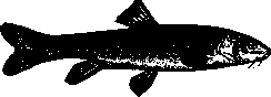
Figure 15. Barbel Barbus barbus. (After Berg.)
upon destruction of the parasite's own body (endotoxins). An example of parasite toxins can be tripanotoxin from various trypanosomes of mammals. Extracts from dried trypanosomes, when injected into mice, cause convulsions and even death in them. Various parasitic worms are undoubtedly toxic. The broad tapeworm can cause malignant anemia; severe intoxication occurs in ankylostomiasis; ascarids possess toxicity. The toxicity of parasitic worms is well detected in experiments with isolated organs. In experiments by Pavlovsky and Dunaeva, an extract from the broad tapeworm (Diphyllobothrium latum) in dilutions from 1:5,000 to 1:100,000,000 in the vast majority of cases caused inhibition of the work of the isolated heart of vagotropic nature (Fig. 16). Their experiments with the effect of extracts of Taenia saginata on the isolated rabbit heart showed a two-phase action - first a brief excitation, then prolonged inhibition of heart activity. The toxin of the unarmed tapeworm should be considered muscular and vagotropic. Experiments
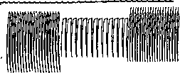
A
B
Figure 16. Effect of extract from the broad tapeworm on the isolated heart: A - normal; B - effect of extract (washing according to B. Pavlovsky and V. Dunaeva). F. Talyzina's experiments with the effect of extract from the broad tapeworm on smooth muscle organs showed an inhibitory effect
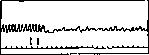
Figure 17. Effect of extract
from the broad tapeworm in a dose of 1:1000 on the rabbit's intestine. (According to Talyzin.) of toxin on the motor function of the isolated intestine (Fig. 17). III. Accidentally poisonous animals may become so either during their life or after death. Some edible mollusks become poisonous during their lifetime. It is indicated that when inhabiting polluted sea water, for example, in the inner harbor, edible mollusks - mussels (Mytilus edulis) (Fig. 18) become poisonous and cause poisoning after being eaten, which can even end in death. The poisonous principle of mussels, according to Brieger, is mylilotoxin (causing curare-like paralysis) and mylilokongestin (Richet), which gives phenomena of anaphylaxis. When mollusks are transferred to clean water, they gradually lose their toxic properties. Some animals may become poisonous when feeding on harmless for themselves but poisonous for other insects. For example, frogs that feed on blister beetles become poisonous to humans (when consumed). The category of accidental animal toxicity includes the laying by bees of the so-called "drunken honey". Its consumption causes intoxication, vomiting, loss of consciousness for up to a day, etc. The cause of the toxicity of honey is not precisely established. It is possible that here the role is played by bees collecting honey from rhododendron flowers (in them
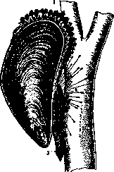
Fig. 18. Mussel - Mytilus edulis: 1 - edge of the mantle; 2 - byssus; 3 - foot. (According to Lang.) glycoside andromedotoxin) or its toxicity depends on careless honey collection, when bees get into it and are crushed there. Finally, accidentally poisonous animals include fish possessing "fish poison". The latter is an exogenous body in relation to the fish and depends on the vital activity of bacteria (see Botulism). - Treatment of poisonings by animal toxins is specific when there are antitoxic sera, or it is of a symptomatic nature.
Related articles
Mentioned in
Cite this page
“Poisonous Animals.” Soviet Medical Encyclopedia. English translation of Bolshaya Meditsinskaya Entsiklopediya, 1st ed. (Moscow, 1928–1936), ed. N. A. Semashko. https://sovietmedicalencyclopedia.pages.dev/article/poisonous-animals/