Counting Chambers
Historical document, translated for reference. It reflects medical knowledge of the 1920s–30s and is not medical advice.
Summary
This article describes the construction and use of counting chambers (hemocytometers), such as the Thoma-Zeiss and Bürker models, used for quantifying blood cells and other microscopic particles. It details the grid systems, dilution techniques using mixers, and the mathematical formulas required to calculate cell concentrations per cubic millimeter.
Encyclopedia article (1928–1936)
COUNTING CHAMBERS, a device allowing for the counting of formed elements of blood and cerebrospinal fluid, bacteria, etc. All counting chambers represent a thick microscope slide, in the middle of which is a depression having a uniform depth in all its parts—0.1 mm, in some counting chambers 0.2 mm (see below). At the bottom of this depression is engraved a grid divided into squares of a specific area (usually 1/400 mm²). A drop of the liquid under investigation, diluted accordingly, is applied to the depression, and since the volume of liquid above each square is precisely known, it is easy to calculate the quantity of formed elements in 1 mm³ from the number of formed elements in one square. One of the most commonly used chambers for counting formed blood elements is the Thoma-Zeiss chamber. Its construction consists of the following: a square glass plate with a round hole in the middle is glued onto a microscope slide. In this hole, a round plate equipped with a grid (Fig. 1) is glued to the microscope slide. This plate is 0.1 mm thinner than the square plate. Thus, if the hole in the plate is covered with a cover slip, the distance between the lower surface of the cover slip and the round plate will equal 0.1 mm. Between the plates, there is a circular groove necessary so that excess liquid is contained in the chamber. Mutually perpendicular
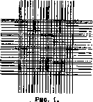
grid lines are located at a distance of 1/20 mm from each other and thus form squares with an area of 1/400 mm². Every fourth line is triple. The intersection of these triple lines forms fields containing 16 small squares. The length of the side of a large square = 0.2 mm (1/20 x 4), and the area = 1/25 mm². In total, there are 16 large fields in this grid. The imperfection of the Thoma chamber lies in the following: filling it with blood is performed before applying the cover slip. Therefore, the chamber may either not be completely filled, and then the depth of the blood layer will be less than 0.1 mm, or the drop will protrude from the chamber, and then precise grinding of the cover slip will be difficult. The grid of the Thoma chamber
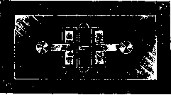
Figure 2.
is arranged in such a way that the large squares are not clearly separated from the small ones and the number of these fields is limited. The disadvantages of the Thoma chamber itself are eliminated in the Bürker chamber, and a number of other grids have been proposed instead of the Thoma grid. Bürker chamber. In this chamber, two elongated support plates B are fixed on a microscope slide (Fig. 2), and between them are glued two counting plates A equipped with a grid (Fig. 4), which, as in the Thoma chamber, are 0.1 mm thinner than plates B. The counting plates are separated by a groove, and their rounded outer ends, as seen in the figure, protrude beyond the ends of plates B. When a cover slip is tightly ground onto plates B, the ends of the counting plates protrude from under the cover slip to the outside. If one now applies a drop of diluted blood to this protruding end of the counting plate, then due to capillarity, the blood will immediately be sucked into the space between the cover slip and the counting plate, i.e., it will precisely occupy the space having a depth of 0.1 mm. This, as well as the ability to perform a control determination thanks to the presence of, as it were, two chambers, constitutes the great advantage of the Bürker chamber. The Bürker chamber is equipped with various grids, the arrangement of which can be seen in Figure 4. The Türk grid (Türk; Figure 3) is a modification of the Thoma grid, as its central part is identical to the Thoma grid; but the bands diverging from this part in a cross shape are equipped only with fields (large squares), and the 4 corners of this grid are completely identical to the Bürker grid (in each corner there are 16 large squares). The total area of the Türk grid = 9 mm². The Bürker grid (Figure 4) represents 9 square fields formed by the intersection of double, Fig. 4
Figure 5. counting in the largest possible number of squares and to most sharply separate large squares from small ones. This is done because it is impossible to limit oneself to counting formed elements in a few squares. The volume of blood in each field is equal to 1/250 mm³ (1/25 x 1/10). For counting erythrocytes, blood is diluted 100 or 200 times.
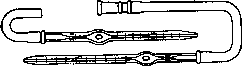
Figure 6.
To determine the number of erythrocytes in 1 mm³, one must multiply their number in one large square by 25,000 (with a 100-fold dilution) or by 50,000 (with a 200-fold dilution); thus, the error increases by the same factor. Therefore, it is necessary to perform the count in a larger number of squares and calculate per 1 mm³ only the average obtained from a large quantity of figures that do not differ significantly from one another. Since there are very few leukocytes in the blood compared to erythrocytes, their count can be conducted only in large squares (fields); hence the importance of a convenient arrangement of large fields in the grids for counting. Erythrocytes are always counted in all 16 small squares. Therefore, it is convenient when small squares are gathered into groups, as is the case in the Türk, Predtechensky, and Neubauer chambers. General methodological instructions (see also Blood). Blood must be diluted to count formed elements, otherwise the formed elements will overlap one another; dilution is achieved by using a mixer (Fig. 6). The lower end of the mixer is drawn out into a capillary; a tube is placed on the upper end, through which blood is sucked up to the mark. In the middle, there is an expansion inside which a glass bead is placed. Blood is sucked up to the 0.5 mark (in the middle of the capillary) or to the 1 mark and then immediately diluted with the diluting fluid. One should beware of blood clotting in the capillary. For counting erythrocytes, a mixer is taken whose ampoule volume (up to the 101 mark) is 100 times greater than the volume of the capillary up to the 1 mark, and for counting leukocytes, a mixer with an ampoule whose volume is 10 times greater than the volume of the capillary. Thus, one can take a dilution of 100 and 200 (if taking blood up to the 0.5 mark) times or, accordingly, for leukocytes, 10 and 20 times. (For staining leukocytes, the composition of diluting fluids, and the method of taking blood, see Blood.) After mixing and shaking the blood with the diluting fluid, 1-2 drops of pure solvent remaining in the capillary are blown out of the mixer, and then a drop of diluted blood is transferred to the counting chamber. The cover slip in both the Thoma chamber and the Bürker chamber (in the latter, beforehand) must be perfectly ground to the support plate. The precision of the grinding is recognized by the appearance of multicolored, so-called Newton's rings between the cover slip and the support plate. Counting is performed, as stated, in a series of squares under a microscope at medium magnification (250x). For Zeiss microscopes, objective C is convenient, for Reichert—6b, for Leitz—5. Corresponding eyepieces—3 (Zeiss), 2 (Reichert), 2-4 (Leitz). The microscope diaphragm is narrowed. A rotating stage is very convenient. Cover slips 0.3-0.5 mm. Counting technique. To avoid counting the same corpuscle twice, it is a rule to count those formed elements that lie on the upper and left lines of a given square, and not to count those that lie on its right and lower lines. It is understood that these elements will then be counted when counting in the square adjacent from below or from the right. The calculation per 1 mm³ occurs as follows: if one large square (containing 16 small squares) contains A erythrocytes, then in 1 mm³ there are 25,000 A with a 100-fold dilution and 50,000 A with a 200-fold dilution (for leukocytes, 2,500 A and 5,000 A respectively). This is understood from the fact that the volume of each large square = 1/250 mm³, and the volume of a small square is equal to 1/4000 mm³. The area of the square and the depth of the counting chamber are engraved on all chambers. When counting leukocytes in the entire chamber, the total area of the grid is taken into account; thus, if N leukocytes are counted in the entire Bürker chamber, then in 1 mm³: x = N * 1 / 0.9, since the area of the entire Bürker grid = 9 mm², and its volume = 0.9 mm³; dilution—20 times. In conclusion, it is necessary to note the necessity of absolute cleanliness of both the chamber and the mixer (they are washed sequentially with water, alcohol, and ether).
G. Kopradya. Besides the original Thoma-Zeiss and Bürker chambers, there are a number of their modifications, consisting mainly in increasing the area of the grid, which is important when counting leukocytes. - Modifications of the Thoma-Zeiss chamber. Zappert counting chamber (Figure 7) - the grid area is 9 times larger than in the Thoma-Zeiss chamber; each side of the rectangle is 3 mm. The grid represents the Thoma-Zeiss grid in the middle, with extensions of these lines extending to the sides, forming 8 more squares equal in size to the central square. Elzholz counting chamber - a further improvement of the previous grid (Fig. 8). The corner squares are divided by additional double lines, which facilitates the counting of leukocytes. The depth of both of the latter chambers is 0.1 mm. Modifications of the Bürker chamber. Klyucharev counting chamber - represents a Bürker chamber with a Predtechensky grid (Fig. 5). Goryaev counting chamber - the same as the previous one, with an expanded Predtechensky grid. - Goryaev-Pappenheim counting chamber represents a Bürker chamber with a Goryaev-Pappenheim grid (Fig. 9). In this grid, which resembles the Predtechensky grid, the size of the undivided large squares has been reduced to facilitate the counting of leukocytes by introducing additional lines. The total number of squares, as in the Predtechensky grid, equals 100, the number of large squares divided into 16 small ones is reduced to 16; the number of large undivided squares is increased to 36. The total area of the grid and the depth are the same as in the Klyucharev chamber. In addition to the two basic types of counting chambers described above, there are more advanced chambers made of solid glass, which eliminates the possibility of the counting plate detaching and the appearance of the grid changing when the balsam gluing it dries. These include: Levy counting chamber; made of solid glass according to the type of the open Bürker chamber with a single (Fig. 10) or double (Fig. 11 and 12) Türk grid (Fig. 3) or Neubauer grid (Figure 13). The latter resembles the Türk grid, but the extreme squares in it are divided not by double, but by single lines. The chamber is filled after placing the cover glass; the drop is sucked into the chamber by capillarity, as in the Bürker chamber. The total surface of the grid is 9 mm2, the depth of the chamber is 0.1 mm. - The Neubauer grid is very convenient and is used
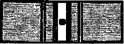
Figure 10.
in chambers of other systems as well. Released by the Zeiss and Leitz firms. - Hausser counting chamber (Hausser; Fig. 14) consists of two parts: 1) a glass part, representing a slide with a chamber, analogous to the Levy chamber, but of a significantly smaller size (Fig. 15 and 16), and 2) a case into which the glass part is inserted (Fig. 17). Special clamps (Figure 18) fix the cover glass to the
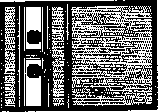
Figure 11.
slide. In the Hausser chamber, Türk, Neubauer, and other grids are used. The latter two chambers are used in America. (In the Arthur Thomas Co. catalog No. 3300 and 3318.) Counting chambers for counting formed elements of cerebrospinal fluid. For this purpose, chambers with a large surface or with a large depth are used. The Fuchs-Rosenthal counting chamber (Fuchs, Rosenthal) is designed according to the type of the Thoma-Zeiss counting chamber, distinguished by a larger grid surface and greater depth. The depth of the Fuchs-Rosenthal chamber is 0.2 mm, the grid surface is 16 mm2, the volume is 3.2 mm3. The grid is divided by double lines into 16 large squares (Figure 19); each large square is divided by single lines into 16 small squares. The chamber is manufactured by the Zeiss firm. - Glaubermann counting chamber represents a modification of the Fuchs-Rosenthal chamber, close in construction to the type of the Bürker chamber; the grid area is increased to 25 mm2. - Dunger counting chamber - construction according to the type of the Thoma-Zeiss chamber. The depth of the chamber is 0.1 mm, the grid area is 50 mm2, the volume is 5 mm3 (Fig. 20). The central part of the grid consists of 9 squares, analogous to the Türk grid; the central part is surrounded by two rows of large squares; the outer row consists of seven large squares (the side of the square = 1 mm, which is why the total area = 49 mm2); to simplify counting, the total area of the grid is brought to 50 mm2 by adding two lines at the top and bottom, 1/7 mm high and 7 mm long, which makes up the missing 1 mm2. Manufactured by the Zeiss firm. The central part of the grid can be used for ordinary blood element counts, the grid as a whole - for counting leukocytes in cases of significant leukopenia, for counting formed elements of cerebrospinal fluid, for counting eosinophils, mast
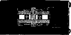
Figure 12.
Figure 13.

Figure 14.
cells, and for cytodiagnostic purposes. - Methodological instructions for counting formed elements of cerebrospinal fluid. For counting in the most common Fuchs-Rosenthal chamber, mixers for white blood cells are used. The liquid serving for dilution (4-5% acetic acid, stained with methyl violet) is drawn up to the 1 mark, and cerebrospinal fluid up to the 11 mark, which achieves a dilution of 1/10. Calculation: if the number of formed elements in the entire chamber is a, then in 1 mm3 there will be X = a * 10 / 3.2 = 3a, which without any special error is replaced by dividing the number obtained during the count of elements in the entire chamber by 3. The count is performed with eyepiece No. 4 and objective No. 5 of a Reichert microscope or corresponding systems of other firms. Counting chambers for bacteria. One can count only emulsions of homogeneous bacteria in a chamber, which do not form clumps, threads, chains, etc.; it is better to do this without any staining, as the dye precipitate interferes with the count. For counting, one can use ordinary chambers, e.g., the Thoma-Zeiss chamber. After thorough shaking, the bacterial emulsion is diluted with physiological saline (0.85% NaCl) using accurately graduated pipettes (Ehrlich) by 10-20 times, if the density of the stock emulsion is approximately 1-2
Figure 15. Figure 16. Figure 17.
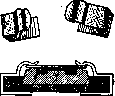
billion. From the diluted emulsion, after thorough shaking, a drop is taken with a capillary pipette and applied in the usual order to the Thoma-Zeiss chamber. No less than 4 large squares are counted. Knowing that the volume of a large square = 1/4000 mm3, the number of microbial bodies in 1 cm3 is determined by the formula X = (n * m * 4000) / q, where q is the number of counted large squares, n is the number of counted bacteria, m is the dilution factor. The count is performed with the largest dry system of the microscope; the use of an oil immersion system is impossible due to the thickness of the cover glass and the depth of the chamber. The depth of ordinary chambers makes counting very difficult, as microbes move due to molecular motion, are arranged in several layers, and do not settle for a long time. To reduce molecular motion, it is recommended to add 0.2-0.5% gum arabic or
Figure 18. 2% peptone solution. - Recently, special, smaller chambers have been used: Thoma counting chamber for bacteria made of solid glass; the construction is analogous to the Levy chamber with a single grid (Fig. 10). The depth of the chamber is 0.02 mm, the grid is of the Zappert type (Fig. 7). The count is performed only in the large squares of the central part of the grid, analogous to the above. When calculating the number of microbes in 1 mm3, the formula must be multiplied by 5, as the volume of this chamber is 5 times less than the volume of the ordinary Thoma-Zeiss chamber and the volume of a large square = 1/1250 mm3. - Troester counting chamber is performed in a darkened field in the eyepiece. For Figure 19. Counting in the counting chamber, the grid is located. For each objective, it is necessary to determine the size of the grid sides using an objective micrometer. The number found for a given combination of lenses and a given length of the microscope tube is a constant value. The calculation is performed according to the formula N = n / (a2 * T), where a is the length of the side of the grid square in millimeters, T is the depth of the chamber, n is the average number of microbes Figure 20. in 1 small square. Helber counting chamber is used in a construction analogous to the Levy chamber (Figure 11) and the Hausser chamber (Fig. 14). The depth of the chamber is 0.02 mm. Neubauer grid (Fig. 13). For the chamber, it is recommended to use especially thin cover glasses - 0.18 mm thick, which makes it possible to use an oil immersion system. The latter is especially convenient in the Hausser chamber due to the fixation of the cover glass with clamps. The chamber is released by the American firm Arthur Thomas Company, Philadelphia. In the catalog Laboratory apparatus and reagents under No. 3305 - Helber chamber in the construction of the Levy chamber; No. 3323 - in the construction of the Hausser chamber; cover glasses - No. 3382. L. Aleksin.
Related articles
Mentioned in
Cite this page
“Counting Chambers.” Soviet Medical Encyclopedia. English translation of Bolshaya Meditsinskaya Entsiklopediya, 1st ed. (Moscow, 1928–1936), ed. N. A. Semashko. https://sovietmedicalencyclopedia.pages.dev/article/counting-chambers/