Graphical Method
Historical document, translated for reference. It reflects medical knowledge of the 1920s–30s and is not medical advice.
Summary
The graphical method is an objective technique for registering physiological phenomena in the human body during both normal and pathological states. It has been instrumental in advancing research on cardiovascular, respiratory, neuromuscular, and other bodily systems through various recording techniques.
Encyclopedia article (1928–1936)
GRAPHICAL METHOD (in experimental and clinical medicine), a method for the objective registration of phenomena in the human body in normal and pathological states. Thanks to the wide application of the G. m., physiology, pharmacology, and various fields of medicine have achieved enormous successes in a short time in the study of the activity of the cardiovascular (cardio-, sphigmo-, plethysmo-, electrocardiography), respiratory (pneumography), neuromuscular (dynamography, ergography), and other systems of the organism. The G. m. has especially firmly established itself among the methods of clinical investigation of nervous patients due to the fact that in the symptomatology of nervous diseases, motor phenomena constitute a large group. By using the method of projecting motor symptoms onto a plane, it is possible to study the functions of various parts of the nervous system under physiological experimental conditions. This method makes it possible to analyze all components of movement (form, rhythm, strength, etc.) and allows changing the conditions of the experiment to account for internal and external influences. The advantage of the G. m. is its objectivity, which makes it possible to exclude the influence of the researcher's personality on the results of observation or experiment. In many cases, when the phenomenon being studied differs in great speed, only the G. m. allows one to investigate its course over time [study of heat production (3,600 times per minute) in a muscle by Hill's myothermic method and others].
The principle of the G. m. is that the movements or changes in potential difference accompanying the phenomenon being studied act on the receiving instrument (cardiograph, non-polarizing electrodes, etc.). This action is transmitted through connecting wires to the recording instrument and with its help is recorded on a tape mounted on the cylinder of a kymograph. The latter instrument (in numerous variants) is an essential component of the G. m. Due to the rotation of the kymograph cylinder, at each subsequent moment a new clean spot on the tape appears under the tip of the recording pen (see Figure 1), and the physiological phenomenon, which always occurs over time, is thus registered in a rectangular coordinate system, in which time is plotted along the abscissa axis. Sometimes for accurate calculations along the abscissa axis, marks are made at certain fractions of a second (most often every 1-0.2-0.01-0.005 sec.) with the help of the so-called Depre marker, which is an electromagnet with a writing pen on the armature. Sometimes the Jacquet time marker clock or a tuning fork is also used for photographic registration. A kymograph with smoked or photosensitive paper wound on its cylinder can be operated at various speeds depending on the speed at which the phenomenon being studied occurs. In the case of rapid phenomena, a plate moving under the influence of a weight or spring (Fallapparat) is used instead of the kymograph cylinder. The design of receiving, transmitting, and recording instruments depends on the nature of the processes being studied. Mechanical transmission. In cases where the phenomenon being studied is associated with movement (muscle contraction, frog heart), a thread, string, or wire is used as the transmission mechanism (myograph, ergograph; see Figure 1). The movements of the string are transmitted to the recording instrument—a lever that records the movement being studied on the kymograph cylinder in an enlarged size. However, mechanical transmission is far from perfect: the string has great inertia and is unsuitable for transmitting rapid movements or over significant distances. More flexible is the air transmission, first proposed by Marey. Changes in the pressure of the air enclosed inside the receiving instrument are transmitted through a column of air in a connecting rubber tube to Marey's recording drum, with the help of which they are recorded on the kymograph. Although air transmission is more sensitive and convenient than mechanical transmission, very rapid phenomena also cannot be registered with its help. The G. m. in its ideal form should make it possible to record phenomena objectively, accurately, and extremely rapidly, and the recording instruments should not distort the picture of the phenomenon being studied.

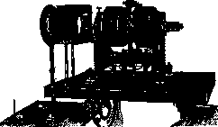
Figure 2. String galvanometer. Photographic recording of string movements. Left - the galvanometer, the meniscus space of which is illuminated by a strong beam of light (the arc lamp on the left is not shown). This convergent beam of light is transformed by a projection eyepiece into a divergent beam, which falls on the front wall of the recording apparatus (box in the upper part of the apparatus shown on the right). Inside the box is a roll of photosensitive paper, unwinding with the help of an electric motor (at the bottom).
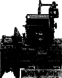
Figure 3. Recording apparatus from the front. Light enters the apparatus through the horizontal slit with divisions shown in the figure. The Jacquet chronograph (on the stand on the right) marks time on the same photosensitive tape, as the shadow of its pen is recorded on the tape in the form of a sine wave. Two transparent circles with numbers (in the middle of the figure) serve for numbering the tapes.
Electric transmission is carried out by an electric current, which has no inertia and has a speed of 300,000 km/sec. If the phenomenon being studied is accompanied by a change in potential difference, then the current transmits this to a mirror or string galvanometer (see Figures 2 and 3). The movements of the string or mirror are further transmitted to photosensitive paper with the help of a light beam, which has no inertia, in contrast to the threads and levers of mechanical transmission. If the phenomenon being studied is not accompanied by the appearance of electricity, then thermocouples and other instruments are used to convert other forms of energy (heat, chemical energy) into electricity and already in the form of electricity to transmit changes in the process being studied to the recording instrument. In newly constructed apparatuses, electricity and light are those transmitters that make it possible to achieve the objective, accurate, and rapid recording of many physiological phenomena. Thanks to this, the graphical method greatly helps the progress of experimental sciences studying humans and animals.
Related articles
Mentioned in
Cite this page
“Graphical Method.” Soviet Medical Encyclopedia. English translation of Bolshaya Meditsinskaya Entsiklopediya, 1st ed. (Moscow, 1928–1936), ed. N. A. Semashko. https://sovietmedicalencyclopedia.pages.dev/article/graphical-method/